Is the animal neutered or not? This question always arises when stray or shelter animals with missing neutering scars and unclear neutering status are presented or older, supposedly neutered animals start showing typical sexual behaviour such as mounting, barking, chasing and increased aggression.
There are various possible causes:
- The animal is not neutered
- The animal is incompletely castrated (ovarian remnant syndrome (ORS), cryptorchidism or residual testicular tissue)
- The animal is castrated but has a sex hormone-producing tumour, e.g. in the adrenal glands (hyperadrenocorticism)
-
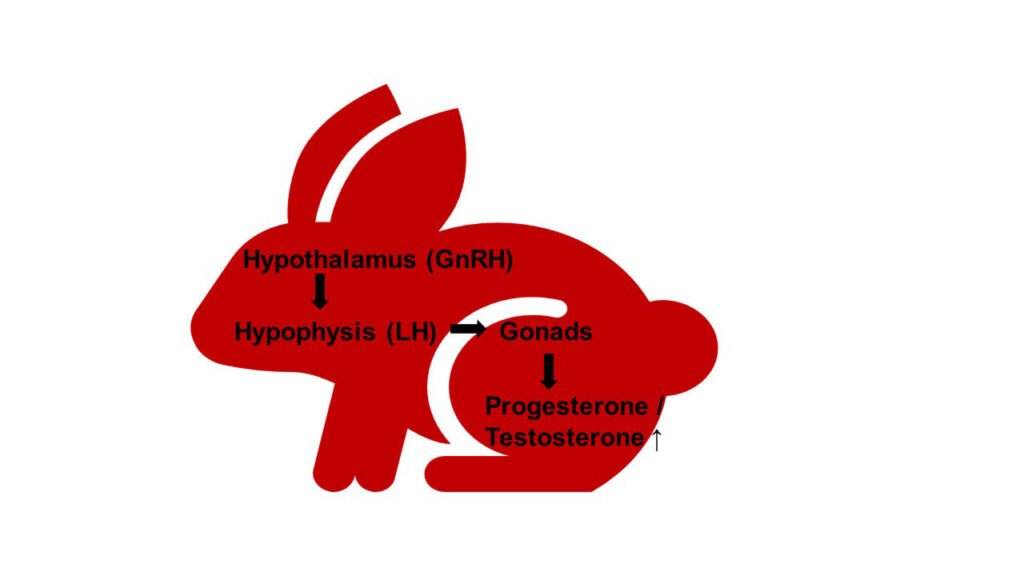
Fig. 1: Schematic representation of the hypothalamic-pituitary-ovarian-testicular axis
Image source: J. Liebscher
It is important to ask questions about possible associations, the prevention of sex organspecific neoplasia (ovarian, uterine and testicular tumours) and their diagnosis as well as the possible treatment of existing neoplasia.
How does laboratory diagnostics help?
Each laboratory test is based on a detailed preliminary report and a thorough clinical examination. If no clear evidence of the neutering status is found here, the blood test helps. In principle, there are three different options, which are more or less suitable depending on the species and age of the animal:
- Basal value determination of classic reproductive hormone concentrations (testosterone, oestradiol, progesterone, 17-OH progesterone and other supplementary steroid hormones)
- HCG stimulation test (with 2 progesterone or testosterone concentration measurements)
- New for rabbits: Determination of the concentration of anti-Müllerian hormone (AMH)
The advantages and disadvantages of the individual tests for different small mammals are discussed below.
1. Basal value determinations of classical reproductive hormone concentrations
In principle, it is possible to determine individual sex hormones in small mammals however, reliable reference values are only available for a few animal species. Typically, the progesterone concentration is measured in females and the testosterone concentration in males.
Basal value determinations are only conclusive with regard to neutering status when hormone concentrations are high.
Low individual values (basal values) are not diagnostically conclusive:
- The animals can be neutered.
- Females may be in anoestrus.
- Hormone production is cyclically at a low point.
The oestradiol concentration is subject to strong fluctuations and is therefore unsuitable for answering the question ” neutered yes/no”. However, it can be used for diagnostic purposes in male animals with suspected oestrogen-producing sertoli cell tumours or castrated ferrets with suspected hyperadrenocorticism.
Androgens and 17-OH-progesterone only play a role in the diagnosis of hyperadrenocorticism in neutered ferrets and can be requested in the package (NNR profile ferrets).
2. HCG stimulation test (with 2 progesterone or testosterone concentration measurements)
For the clear detection of gonadal tissue, an HCG stimulation test with the concentration measurement of progesterone in females or testosterone in males is useful. GnRH stimulates the secretion of LH from the anterior pituitary gland. LH in turn stimulates the production of progesterone in the ovaries and testosterone in the Leydig cells (Figure 1). Buserelin preparations (GnRH analogue, Receptal®, Buserelin®, Veterelin®) are currently approved for small mammals – and here only for rabbits. However, in vitro studies have shown that buserelin reduces progesterone production in the mid and late luteal phase (Zerani et al. 2010) and can therefore lead to falsely low results. HCG preparations (Ovogest®, Suigonan®) are therefore used more frequently. They are not authorised for use in small mammals and must be rededicated accordingly, but have been tried and tested many times and are easy to use.
Performance and interpretation of the stimulation test in rabbits are described in Table 1. No specific tests have yet been described in the literature for other small mammals, but transfer is very possible.
Determination of the concentration of anti-Müllerian hormone (AMH) in rabbits
A good alternative to the HCG stimulation test in rabbits is the AMH concentration determination. In dogs and cats, AMH is now routinely used to differentiate between neutered/unneutered and to diagnose ORS or cryptorchidism. AMH is also suitable for the diagnosis of a granulosa cell tumour in mares, female dogs and cows as well as for the diagnosis of a sertoli cell tumour in male dogs. In horses, AMH is also used to diagnose cryptorchidism (Böhmer 2023).
Table 1: HCG stimulation test for the diagnosis of gonadal tissue in rabbits ( modelled after Geyer 2015, Schützenhofer 2011)
| Male | Female | |
| Procedure: | ||
| 1st blood sampling (basal value): | Determination of testosterone | Determination of progesterone |
| Injection of: | 0.8 μg buserelin (e.g. Receptal®) or 100- 250 IU/animal HCG (e.g. Ovogest®) i. m. | |
| 2nd blood sampling (stimulation value): | Blood sampling after 1 hour | Blood sampling after 5 – 7 days |
| Interpretation of stimulation values: | Testosterone | Progesterone |
| Hormone-producing tissue present (unneutered, ORS) | > 1 ng/ml | > 4 ng/ml |
| Questionable result | 0.1 – 1 ng/ml | 2 – 4 ng/ml |
| No hormone-producing tissue (neutered) | < 0.1 ng/ml | < 2 ng/ml |
AMH is a dimetric glycoprotein that is involved in foetal sexual differentiation. In males, it leads to the suppression of the development of Müllerian ducts. At the same time, the epididymis, vas deferens and seminal vesicle glands differentiate from the Wolffian ducts under the influence of testosterone. In the female, this inhibition by AMH does not take place and the Müllerian ducts then develop into the oviduct, uterus, cervix and cranial vagina.
In sexually mature animals, AMH is produced exclusively in the granulosa cells of the ovaries and in the Sertoli cells of the testicles, regardless of the menstrual cycle (Böhmer 2023).
New publications show that the tests used to determine AMH concentrations in other animal species are also suitable for measuring concentrations in rabbits (Böhmer et al. 2022). Böhmer and his colleagues (2022) used Laboklin to analyse AMH concentrations by chemiluminescence assay (CLIA) in 64 intact and 22 spayed adult female rabbits for the differentiation of spayed/unspayed and AMH concentration in relation to pseudopregnancy and number of follicles (Böhmer et al. 2022).
The progesterone concentration was also measured to determine whether the females were pseudopregnant (< 2 ng/ml: follicular phase, not pseudopregnant, > 2 ng/ml: luteal phase, pseudopregnant). All spayed rabbits showed AMH concentrations < 0.07 ng/ml, differed highly significantly (p < 0.001) from those of intact female rabbits and the value ranges did not overlap (Table 2). There was no significant difference in the follicular and luteal phase (p < 0.951).
As part of further internal Laboklin studies (2023), similar data was measured in 33 male castrated rabbits with the identical device using CLIA (Table 2). A preliminary reference range of < 0.07 ng/ml was established for the male castrated rabbit.
Table 2: AMH concentrations in rabbits (CLIA; female animals, according to Böhmer et al. 2022; male animals, unpublished data from Laboklin)
| Neutering statuss | Quantity (n) | Mean ± standard deviation (ng/ml) | Median (ng/ml) | Range (ng/ml) |
| female spayed | 22 | 0.05 ± 0.04 | 0.06 | 0.01 – 0.23 |
| female intact | 64 | 1.67 ± 0.64 | 1.53 | 0.77 – 3.36 |
|
Female rabbit: AMH < 0.07 ng/ml → spayed |
||||
| male castrated | 33 | 0.04 ± 0.03 | 0.03 | 0.01 – 0.12 |
| male intact | 11 | 14.00 ± 7.83 | 12.94 | 3.76 – 22.96 |
|
Male rabbit: AMH < 0.07 ng/ml → castrated |
||||
The results are consistent with those of other studies (Schwarze 2023), although different devices and test methods were used there.
Differences between intact and cryptorchid males have not yet been investigated. In dogs, calves and stallions, cryptorchids have higher AMH concentrations than intact ones due to immature Sertoli cells and/or lack of suppression by testosterone (Böhmer 2023).
AMH determination is therefore suitable for checking the neutering status of both female and male rabbits. The advantage is that blood is taken once without injection and the result is therefore available quickly. The disadvantage is the sensitivity of the sample, which requires the submission of cooled, centrifuged and pipetted serum (at least 200 μl). AMH concentrations above 0.07 ng/ml are indicative of gonadal tissue.
Further studies are needed on the applicability of AMH measurements in other small mammals and in the context of ORS and granulosa cell/ sertoli cell tumour diagnostics as well as in the field of hyperadrenocorticism in small mammals.
Conclusion
The gold standard for differentiating between neutered and unneutered small mammals is the HCG stimulation test with 2 progesterone/ testosterone measurements. Single measurements are only conclusive for unneutered animals if the concentrations are high. In rabbits, the AMH determination is a good alternative.
Jana Liebscher Dr. Jutta Hein
Range of services
- Testosterone
- Progesterone
- HCG stimulation test
- Anti-Müllerian hormone (rabbit)


