In dogs, tumours mostly occur at the age of 9 – 12 years. However, some young dogs (1 – 3 years) also develop neoplastic masses. So far, only a few studies have been conducted on neoplasms in dogs up to 12 months of age (Kessler & v. Bomhard 1997; Schmidt et al. 2010). Most of these neoplasms (almost 90%) were benign canine cutaneous histiocytomas, but some cases of malignant tumours (sarcomas, lymphomas, carcinomas) were described as well (Kessler & v. Bomhard 1997, Schmidt et al. 2010).
LABOKLIN’s sample material 2016 – 2019
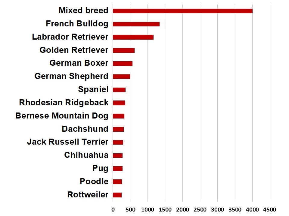
As part of a large interdisciplinary joint research project (FORTiTHer), 170,000 data sets obtained from routine histopathological examinations (2016 – 2019) were analysed. For the results presented here, we evaluated data of dogs up to 3 years of age, for which breed and age were indicated in the clinical history and where the quality of the sample was sufficient for diagnosis (n=18,389). Overall, 225 breeds were included; the 15 most common ones are shown in Fig. 1.
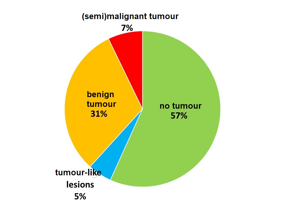
Non-tumorous changes (inflammation, cysts, hyperplasia, degenerative lesions) were detected in more than half (57%) of the submitted samples.
Polyps, dysplasia or epulides, for example, were classified as tumour-like lesions (5%).
Tumours were found in 38% of all submitted samples (Fig. 2).
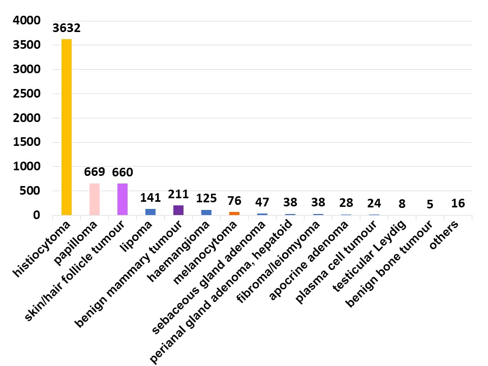
Within the group of tumours, 81% were benign (mainly histiocytomas, papillomas, benign mammary tumours, hair follicle tumours, lipomas, Fig. 3).
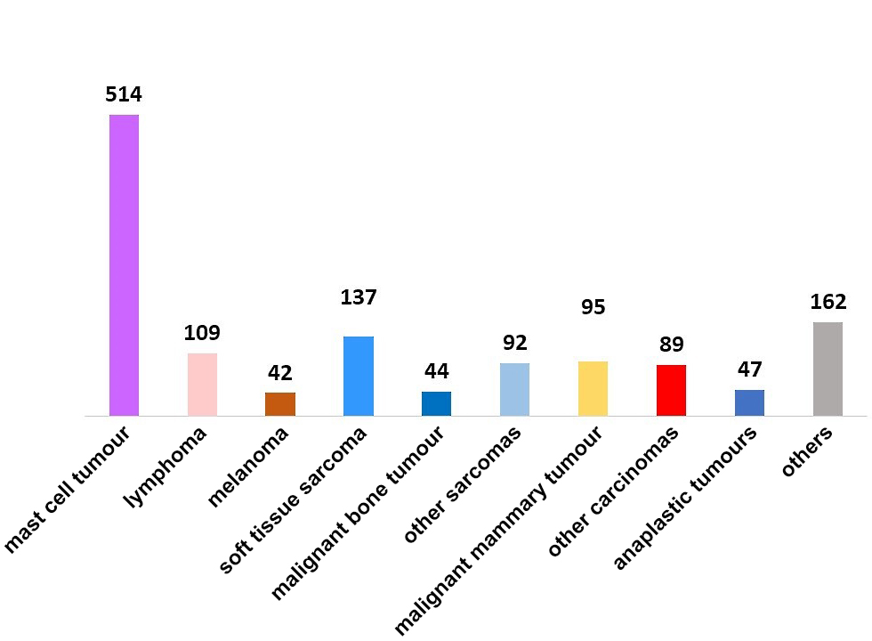
However, this also means that in dogs under 3 years of age, 19% of all tumours submitted were (semi)malignant (Fig. 4)!
Below, a few selected tumours are presented as an example.
Canine cutane histiocytoma
Canine cutaneous histiocytomas are, by far, the most common tumours in young dogs (Fig. 3). Histiocytomas are benign and undergo spontaneous regression in many cases. They are diagnosed by cytology or histology.
Mast cell tumours
Generally, it must always be presumed that a mast cell tumour has malignant potential. In dogs up to 3 years of age, they were the most frequent (semi)malignant tumours (39%, Fig. 4).
Often, the initial diagnosis of mast cell tumours is made cytologically. If possible, the following excision is done paying particular attention to the resection margins and with tumour bed biopsies being collected. Grading and examination of the tumour margins are done histologically.
In our sample material of dogs up to 3 years of age, 44.6% of the mast cell tumours were classified as grade I, 52.7% as grade II and 2.7% as grade III according to Patnaik et al. (1984) (or 97.3% low-grade and 2.7% high-grade, respectively, according to Kuipel et al. 2011).
Lymphomas
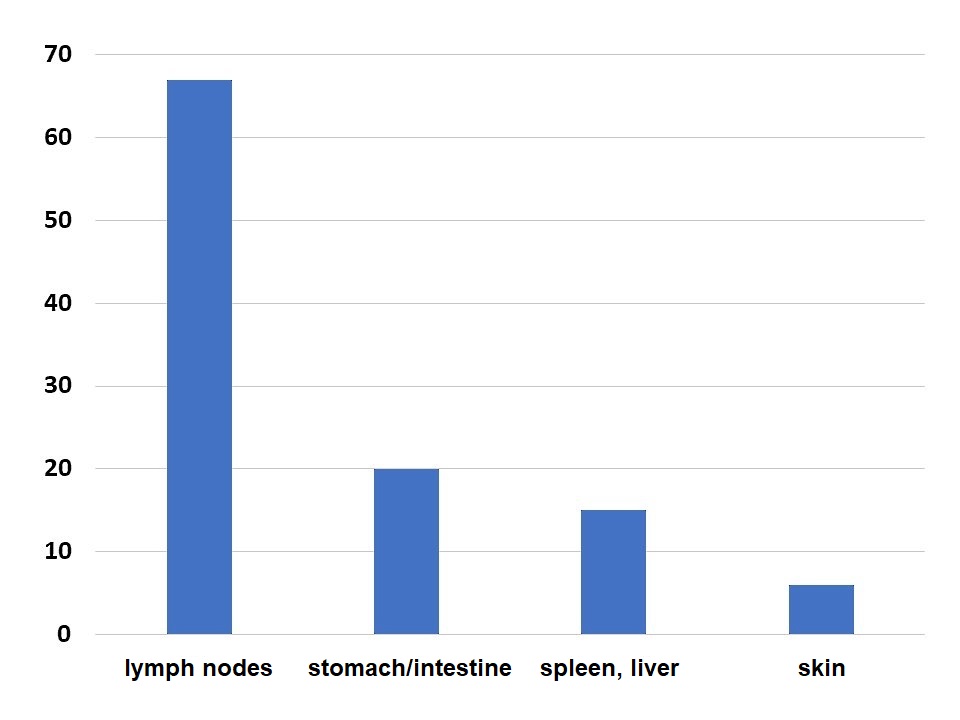
Lymphomas account for 8% of the (semi)malignant tumours in our sample material. The submitted samples mainly originated from the lymph nodes. However, the gastrointestinal tract, the internal organs and the skin were affected as well (Fig. 5).
Lymphomas are usually diagnosed by cytology and/or histology. Further tests (e.g. clonality analysis, immunohistology, immunotyping) provide for a more accurate characterisation.
Soft tissue sarcomas
In our sample material of dogs up to 3 years of age, soft tissue sarcomas were relatively common making up 10% of the (semi)malignant tumours.
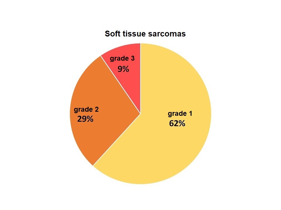
Similar to mast cell tumours, a cytological diagnosis of soft tissue sarcomas can be helpful in planning the surgical procedure. Grading (McSporran et al. 2009) is then done histologically. Two thirds of the soft tissue sarcomas in our sample material were diagnosed as grade 1 (Fig. 6).
Bone tumours
A total of 199 samples from bones were sent in, of which 41% showed inflammation, 23% tumours and 18% callus formation. Nearly all bone tumours (91%) were malignant (osteosarcomas). It is interesting to note that the 46 osteosarcomas primarily came from Labradors (n=7), Golden Retrievers (n=5) and Rhodesian Ridgebacks (n=5), but only one Great Dane was included.
Bone tumours are therefore an important differential diagnosis in young animals, too.
Diagnosis is made by cytology or by histology with a representative sample being of utmost importance. In 11% of the submitted bone samples, the samples were not representative and no morphological correlate to the clinical or radiological changes could be found.
Mammary tumours
In our sample material, 69% of the mammary tumours in dogs up to 3 years of age (n=306) were benign.
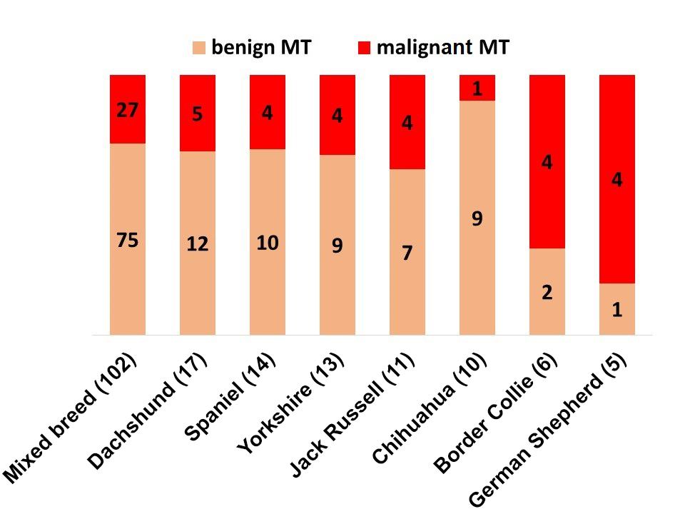
But also simple and complex carcinomas (28%) as well as malignant mixed tumours (3%) were diagnosed. The breed distribution can be seen in Fig. 7.
- Fig. 1: Distribution of the 15 most common dog breeds (≤ 3 years old) in the histological specimens submitted to LABOKLIN (2016 – 2019).
- Fig. 2: Percentage of the diagnoses “no tumour”, “tumour-like lesions” and “benign tumours” or “(semi)malignant tumours” in dogs ≤ 3 years of age in the LABOKLIN sample material (2016 – 2019)
- Fig. 3: Frequency distribution of benign tumours in dogs up to 3 years of age at LABOKLIN (2016 – 2019)
- Fig. 4: Frequency distribution of (semi)malignant tumours in dogs ≤ 3 years of age at LABOKLIN (2016 – 2019)
- Fig. 5: Frequency of organ systems in which lymphomas in dogs up to 3 years of age (n=109) were histologically diagnosed
- Fig. 6: Grading of soft tissue sarcomas (n=136) in dogs up to 3 years of age according to McSporran et al. 2009
- Fig. 7: Breed distribution of dogs up to 3 years of age with mammary tumours (MT) (absolute numbers)
Carcinomas
In our sample material, carcinomas outside the mamma (n=89) originated from a wide variety of organs: mucosa/skin (19 squamous cell carcinomas, 16 apocrine carcinomas, 5 others), urinary tract (n=9), thyroid gland (n=3), pancreas (n=3), anal sac (n=3), liver (n=2), salivary gland (n=2), lung (n=1), ovary (n=1). In 23 cases, the origin of the carcinoma could not be identified/was not indicated. The studies by Kessler & v. Bomhard 1997 and Schmidt et al. 2010 also describe the occurrence of such carcinomas in dogs up to 12 months of age. This proves that the occurrence of highly malignant carcinomas is possible even in such young animals. Diagnosis is usually made by histology or, less frequently, by cytology.
Other tumours
Furthermore, melanocytic tumours (n=111), transmissible venereal tumours (n=60) as well as neoplasms of the ovaries (n=31), testicles (n=35) and dental germs (n=18) were also diagnosed in the young dogs of this study.
Conclusion
In summary, most tumours in young dogs are benign. Histiocytoma is the most common diagnosis.
Nevertheless, even in young animals, almost 20% of all tumours submitted were classified as (semi)malignant, which sometimes means an unfavourable prognosis. Among them, mast cell tumours, lymphomas, sarcomas and various carcinomas played the largest role.
Cytological and histopathological examinations are therefore of great prognostic and therapeutic importance for the differentiation of inflammatory, degenerative, dysplastic, traumatic and neoplastic changes, even in young dogs. Immunohistochemical methods can provide additional information relevant for treatment (e.g. lymphoma typing, differentiation of mast cell tumours).
PD Dr. Heike Aupperle-Lellbach
