Haematology
Urinary crystals in pets

Bilirubin crystalluria

Bilirubin may crystallize in urine to form yellowish-red or reddish-brown needles or granules (Fig. 1). Bilirubin crystals can be observed in highly concentrated urine from clinically unremarkable dogs. If they are found in large numbers in serial urine samples, this suggests a disorder of bilirubin metabolism.

Calcium carbonate crystalluria

Calcium carbonate may crystallize in the urine of horses, rabbits, guinea pigs and goats, forming large, yellow-brown or colorless spheres with radial streaks, or smaller crystals with round, ovoid, or dumbbell-like shapes. Calcium carbonate crystals have not been found in urine from dogs or cats. Dumbbell-shaped crystals found in dog or cat urine are more likely to be calcium monohydrate than calcium carbonate.

Calcium phosphate crystalluria

There are many different types of calcium phosphate crystals. They seem to be described differently as amorphous phosphates and calcium phosphates. With the exception of bruschite, calcium phosphate crystals tend to form in alkaline urine. Under the light microscope, amorphous phosphates resemble amorphous urates (Fig. 18-21). However, amorphous phosphates typically form in alkaline urine and dissolve in acetic acid. In contrast, urates often have a yellow granular appearance and do not dissolve in acetic acid, but do dissolve in alkalis at 60 °C.

Scanning electron micrographs of amorphous phosphates found in human urine revealed that they normally appear spherical but can also take on ring or cast shapes. Only in dogs have we observed the spherical habit of calcium phosphate. Calcium phosphate may also form long, thin colorless prisms, sometimes with pointed ends. These crystals can cluster together in rosettes or appear as needles. Calcium phosphate can also precipitate as elongated lath-shaped bruschite crystals in acidic urine (Fig. 22-23). It has been considered that when calcium phosphate precipitates from an aqueous highly supersaturated solution of pH > 7, initially amorphous crystals appear. The amorphous precipitate can slowly transform into a stable crystalline precipitate by dissolution, conversion and crystal growth. The rate of this transformation is pH dependent because the stability of amorphous phosphates is enhanced at high pH.

Interpretation

Caution must be used in interpreting amorphous crystals discovered under the light microscope, as they form from a variety of crystals, including calcium phosphate, ammonium urate, and xanthine. In our experience, large amounts of presumptive calcium phosphate crystals were observed in clinically unremarkable dogs, dogs with consistently alkaline urine, dogs with calcium phosphate urinary stones, and dogs with urinary stones that were mixed calcium phosphate and calcium oxalate. Small amounts of calcium phosphate crystals may occur in association with infection-related crystalluria.

Cholesterol crystalluria

Cholesterol crystals typically appear as large, flat rectangular platelets with a characteristic corner notch (Fig. 24). Under the light microscope, they are colorless and transparent; under the polarizing microscope, a variety of bright colors are typically observed.InterpretationIn humans, cholesterol crystals have been described to be associated with extensive tissue damage, nephrotic syndrome and chyluria. Veterinary experience with them is too limited to formulate meaningful generalizations. In any case, they were observed in clinically unremarkable dogs.

Cystine crystalluria

Cystine crystals are colorless and have a characteristic hexagonal shape (benzene ring), with sides of equal or unequal length (Fig. 25-26).They can occur individually, but usually accumulate into layers. They are easier to detect when the light intensity is reduced, because they are thin. Cystine crystals form most frequently in concentrated acidic urine. When markedly alkaline urine is produced as a result of infection or contamination from urase-forming bacteria, this can lead to dissolution of cystine crystals. The addition of glacial acetic acid followed by cooling and centrifugation can promote the detection of typical crystals in alkaline urine. Cystine crystals are insoluble in acetic acid, alcohol, acetone, ether and boiling water. They dissolve in ammonia and hydrochloric acid.InterpretationCystine crystalluria is not an insignificant finding. Cystine stones can develop in dogs and cats with the metabolic disorder cystinuria, but not all patients with cystinuria form cystine urinary stones. (See also discussion of magnesium phosphate and uric acid crystalluria for details on differentiating cystine crystals from struvite or uric acid crystals).Some medications excreted in the urine may form crystals. Perhaps the best known drug-induced crystalluria in dogs and cats is that which occurs with sulfonamide use. Sulfonamides may precipitate in urine in characteristic bundles of transparent or brownish needles, usually eccentrically bundled (Fig. 27-29). They may also appear as amorphous crystals or spheres with radial striations (Fig. 30). A positive lignin test reinforces the diagnosis of sulfonamide crystalluria.In humans, leucine crystals indicate serious liver disease. The significance of leucine crystals in dogs has not been extensively researched to date. (See also: Cystinuria in the Newfoundland)

Leucine crystalluria

Leucine crystals typically appear as large yellow or brown spheres with radial concentric layering (Fig. 31). However, such spherical formations may not consist of pure leucine, as it has been described that pure leucine forms crystals resembling hexagonal platelets.In our experience, struvite crystals can be observed in dogs and cats that have 1. are clinically unremarkable, 2. have infection-related struvite urinary stones, 3. have sterile struvitharn stones, 4. Have non-struvitharn stones, 5. Have urinary stones with mixed composition (z.B. a core of calcium oxalate with a shell of struvite) or 6. Urinary tract diseases without urinary stones have.

Struvite crystalluria

Magnesium ammonium phosphate (struvite) crystals typically occur as colorless, orthorhombic (d.h. with three unequal axes intersecting at right angles) coffin-like prisms. They often have three to six or more sides and often have blunt ends (Fig. 32-39). Six- to eight-sided struvite crystals in cats are sometimes mistaken for cystine crystals (Fig. 40-41). However, in contrast to cystine, they occur together with other forms of struvite and dissolve immediately after acidification with dilute acetic acid. Occasionally struvite crystals accumulate to form fern-like structures. The sharp outlines of struvite crystals, characteristic of fresh urine, im can become feathery or moth-eaten during dissolution.InterpretationStruvite crystals commonly occur in dogs and occasionally in cats, along with free ammonia produced during urea microbial hydrolysis triggered by urease. Struvite crystals also occur frequently in cats and occasionally in dogs in the absence of detectable urease. In this case, the ammonia component of struvite is probably generated by the renal tubule. Composite urinary stones containing varying amounts of sulfonamides have been found in dogs and cats. Other types of drug-induced crystalluria have been described only in humans. X-ray contrast agents such as Urografin® (Schering) and Uromiro® (Heyden) may precipitate in urine as pleomorphic needles that occur singly or in bundles. Ampicillin may precipitate in acidic urine as thin colorless needles. Primidone may precipitate as hexagonal platelets resembling calcium oxalate monohydrate. Ciprofloxacin may precipitate in alkaline urine as bundles with eccentric binding.

Urates: ammonium and amorphous urate crystalluria

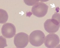
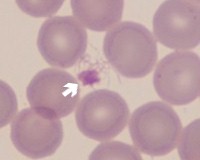
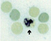
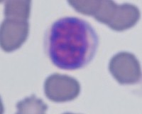
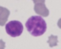
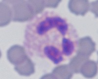
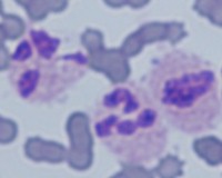
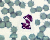
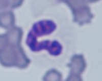
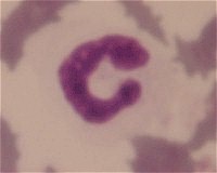
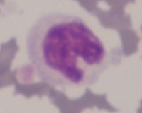
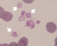
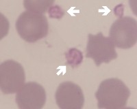
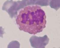
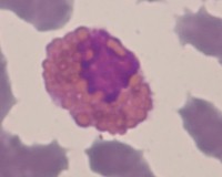
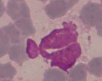
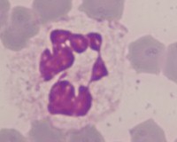
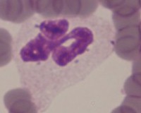
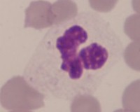
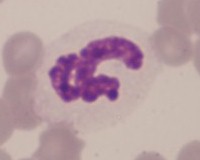
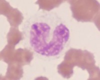
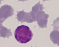
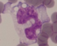
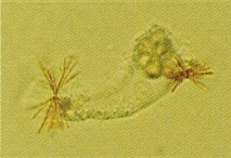
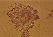
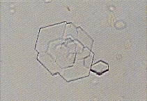
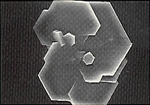
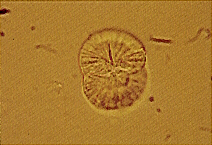
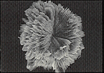
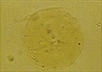
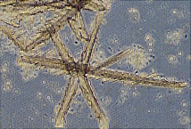
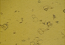
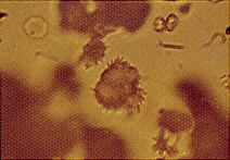
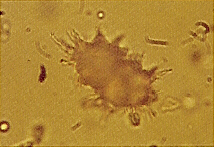
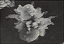
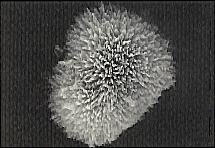
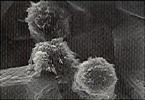
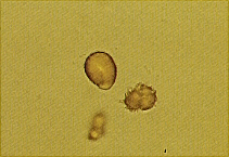
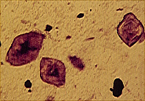
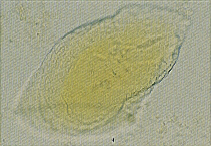
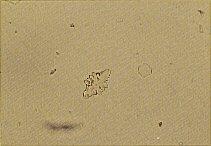
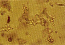
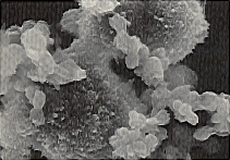
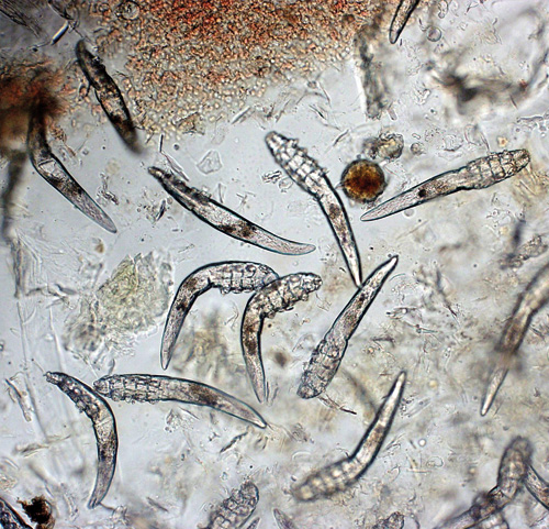
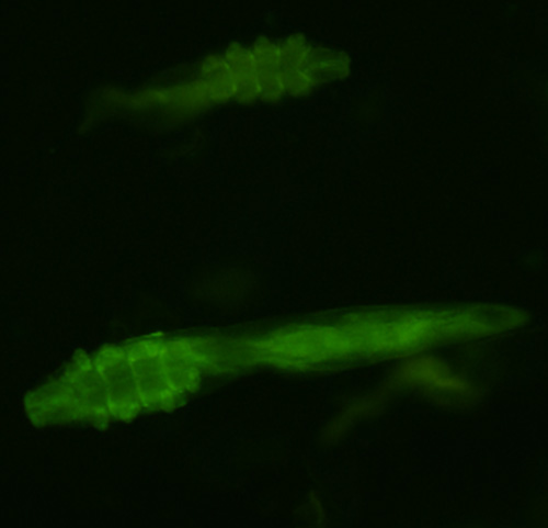
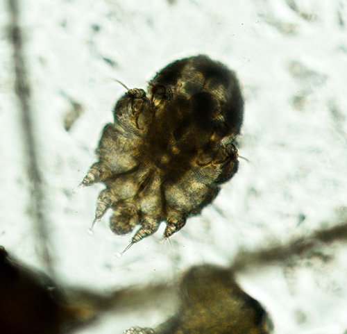
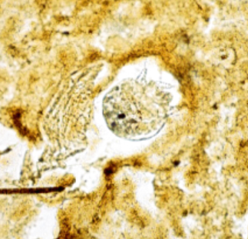
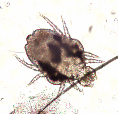
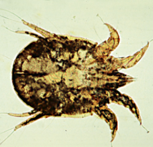
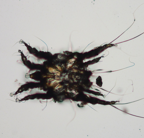
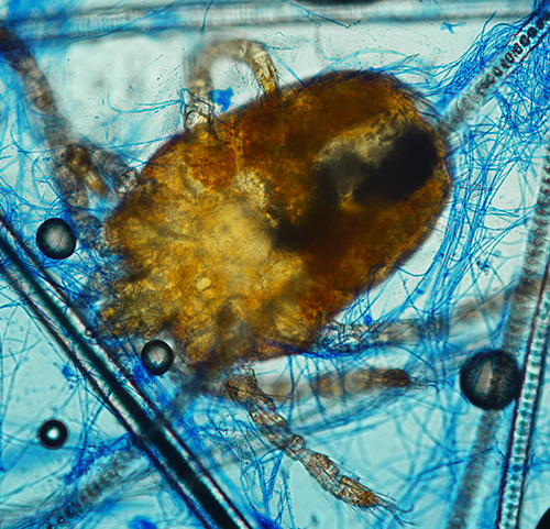
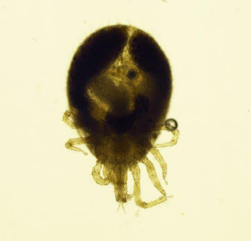
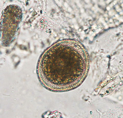
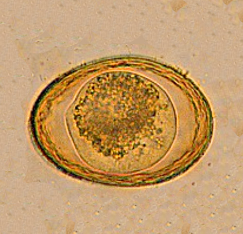
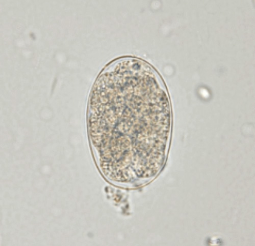
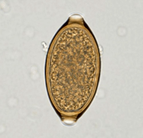
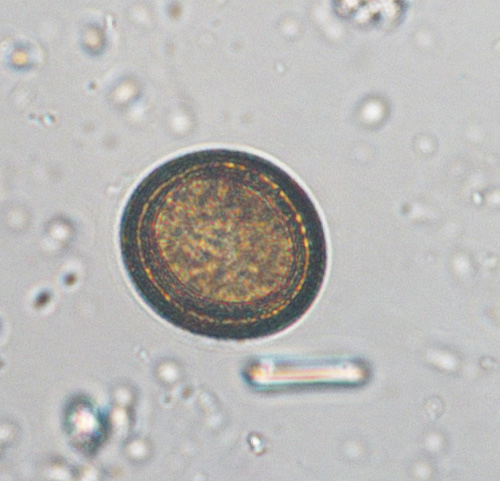
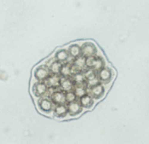
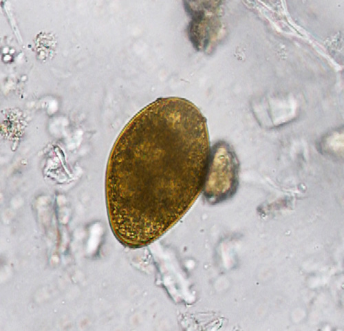
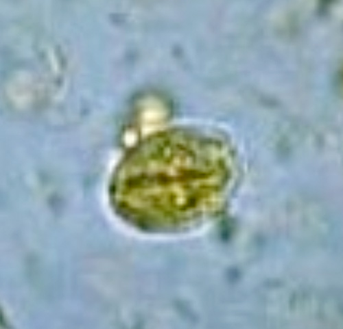
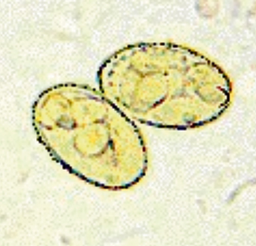
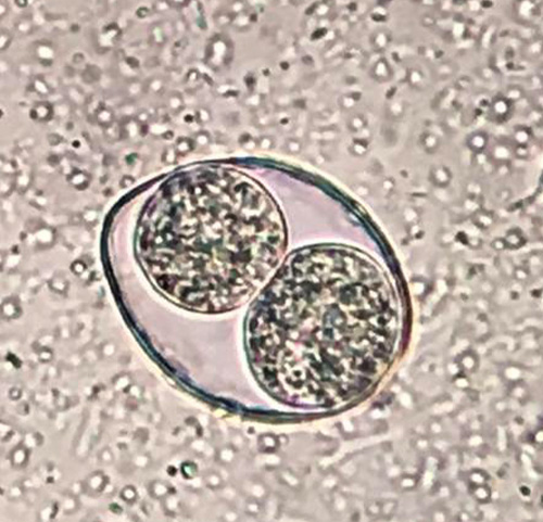
Ammonium urate (also called ammonium biurate) crystals are commonly observed in slightly acidic, neutral and alkaline urine. They are usually brown or yellow-brown and may form globules or spherical bodies with long irregular projections (called datura) (Fig.43-51).

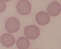
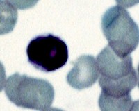
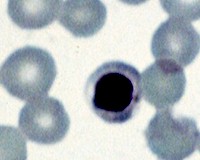
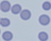
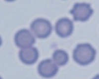
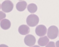
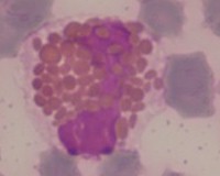
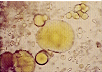
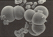
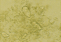
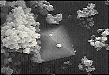
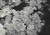
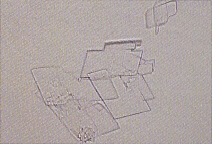
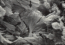
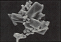
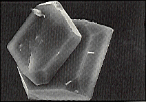
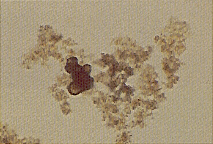
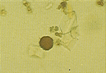
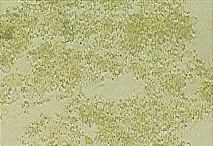
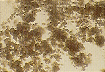
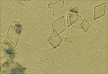
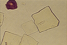
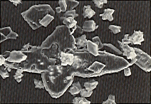
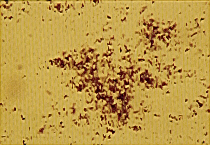
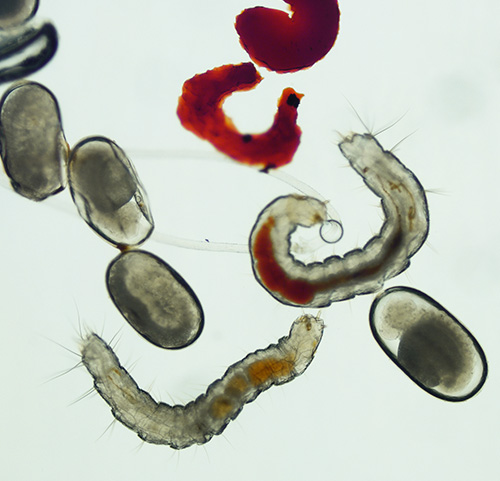
Sodium, potassium, magnesium and calcium urate salts can precipitate in amorphous form in acidic urine (so-called amorphous urates). They can resemble amorphous phosphates (Fig. 49), but dissolve in alkaline environment. When the amorphous crystals grow (Fig. 47-51) they develop a characteristic yellow or yellow-brown color.

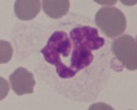
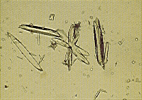
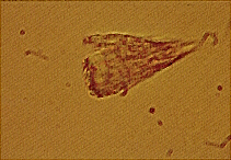
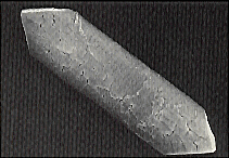
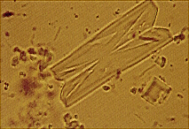
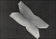
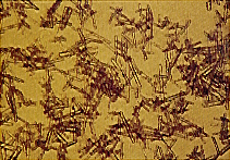
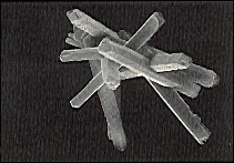
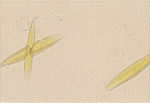
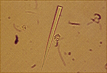
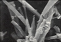
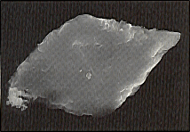
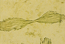
Sodium urate may also precipitate as colorless or yellowish needles, or as slender prisms that appear in bundles or clusters(Fig. 52-56).

Ammonium urate and amorphous urate crystals are not soluble in acetic acid. however, the addition of 10% acetic acid to urine sediment containing these crystals often results in the appearance of uric acid and sometimes sodium urate crystals. (See discussion of uric acid crystalluria for details.) The addition of acetic acid to amorphous phosphate crystals leads to their rapid dissolution, whereas they persist in alkaline urinary sediment.

Interpretation

Ammonium urate and amorphous urates may be present in clinically unremarkable dogs and cats, but they are not common. They are often observed in dogs with portal anoinalia with or without concomitant aminonium urate stones. They are also commonly found in Dalmatians, English Bulldogs, and other dogs and cats with ammonium urate urinary stones caused by conditions other than portal vein abnormalities.

Uric acid crystalluria

Uric acid crystals are often yellow or yellow-brown and can occur in a variety of shapes. The most typical are diamond-shaped or rhombic platelets, which may contain concentric rings (Fig. 57-61). They may also occur as rosettes composed of aggregations of many uric acid crystals. Occasionally, uric acid crystals form rhomboid platelets with one or more paired lateral projections (Figs. 62-63). Less commonly, they appear as six-sided crystals similar to cystine. However, the six-sided crystals occur together with typical diamond or rhombic shapes. Uric acid crystals are soluble in sodium hydroxide solution but insoluble in alcohol, hydrochloric and acetic acid.

Interpretation
Although common in humans, naturally occurring uric acid crystalluria is uncommon in dogs and cats. When noted, the crystals have the same significance as described for ammonium and amorphous urates. Uric acid crystals form rapidly after the addition of 10% acetic acid to canine and feline urine sediment containing amorphous urate or ammonium urate crystals. Sodium urate crystals may also occur. It is often necessary to expose the uric acid crystals to the acetic acid for 20-30 minutes before they become visible. They can grow to considerable size if kept overnight in a covered petri dish moistened with a sponge soaked in water.

Xanthine crystalluria

Xanthine may precipitate in acidic urine in an amorphous form especially in dogs and cats which were given allopurinol. Amorphous xanthine crystals resemble to amorphous urate crystals (Fig. 64-66). The detection of xanthine crystals allows to presume an excessive dose of allopurinol related to the amount of purine predecessor in the food. Xanthine crystalluria in humans has been described in accordance to a hereditary xanthinuria.

Parasites