Proliferative lesions of the oral cavity are regularly seen in both dogs and cats. In contrast to nodules in the skin, which are easily visible, proliferative lesions in the oral cavity often go unnoticed for a long period. Once they reach a certain size, secondary changes like increased salivation or a decreased food-intake indicate presence of a lesion in the oral cavity. A regular and thorough inspection of the oral cavity is therefore important.
In addition to inflammatory lesions, various epithelial, mesenchymal, melanocytic or round cell neoplasia can occur in the oral cavity – similar to other locations and organ systems. However, odontogenic tumours are unique to the oral cavity and do not occur elsewhere in the body. They originate from the various components of the teeth, periodontal apparatus and surrounding stroma. Based on the cell of origin, currently a rather descriptive nomenclature for odontogenic tumours is used, which can be quite long and complicated. The nomenclature is based on the human WHO classification of odontogenic tumours, even though this classification is not always directly applicable to the veterinary odontogenic counterparts. Based on comparative pathology and continuing extending knowledge, the nomenclature has been subject to a constant change in the last decades. The unique features of a selection of four odontogenic proliferative lesions are discussed below.
Canine acanthomatous ameloblastoma
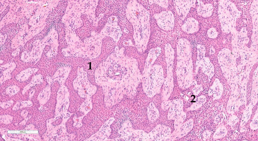
Canine acanthomatous ameloblastomas (CAA) (Figure 2) are unique to dogs, and are formerly known as acanthomatous epulis or peripheral ameloblastoma. It is the most common odontogenic tumour in dogs. CAA can occur anywhere in the oral cavity adjacent to teeth, but in about 50% of the cases the rostral mandible is affected. Especially medium to large breed dogs are reported to have CAA, although all breeds can be affected.
In general, CAA is considered as a benign neoplasm, since metastases have not been reported so far. However, despite being a rather benign lesion, CAA can show highly variable biological behavior, varying from slow growth and minimal local invasion, to rapid and extensive local invasive growth with high tropism for local bone invasion. Especially predominantly intra-osseous CAA are associated with highly aggressive biological behavior. These tumours can therefore cause marked destruction and osteolysis of the underlying mandibular or maxillary bone, with additional tooth loss, which can be identified on radiographs.
-
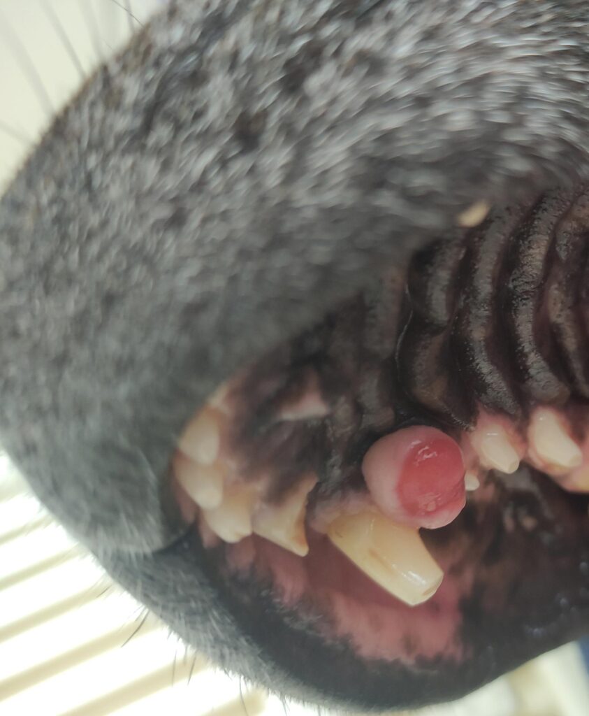
Fig. 1: Accessory formation in the oral cavity of a dog, medial
Photo credit: Laboklin
-
Fig. 2: Histological picture of a CAA, dog. 1) Predominant odontogenic epithelium. 2) Early intraepithelial cyst formation. HE stain, 10x obj.
Photo credit: Laboklin
-
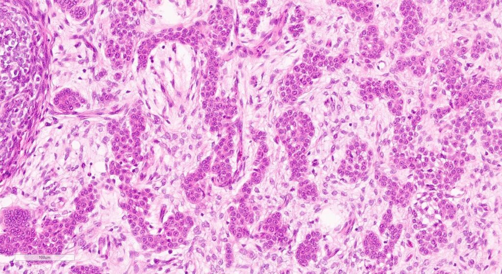
Fig. 3: FIOT, cat. HE staining, 20x obj.
Photo credit: Laboklin
Although the exact cell of origin remains unclear, the most current believe is that CAA rather arises from odontogenic epithelium, instead of oral mucosal epithelium. Immunohistochemical studies were unfortunately not successful to confirm a definitive cell of origin.
Although CAA have a highly variable macroscopical appearance, they appear rather uniform on histology, independent from the biological behavior (slow-progressive versus aggressive invasive growth), in which the odontogenic epithelial component is uniform and predominant. Intra-epithelial cyst formation due to degeneration is possible. The variable intra-epithelial keratinization can make the differentiation between CAA and squamous cell carcinoma challenging on the microscopical level, to sometimes even impossible. In conclusion, besides histological examination, additional diagnostic imaging (radiographs, computed tomography) is a valuable tool for both treatment planning and prognosis prediction. In general, en bloc surgical resection with a tumour free margin of 2 centimeter is recommended. With free margins, the prognosis is good and recurrence rarely occurs.
Fibromatous epulis of periodontal ligament origin / peripheral odontogenic fibroma
Fibromatous epulis of periodontal ligament origin/peripheral odontogenic fibroma (FEPLO/POF), also known as ‘epulis’ or ‘fibromatous/ossifying epulis’, is another very common gingival lesion in dogs (and uncommon in cats) of any age. This lesion can be focal or multifocal and can occur anywhere on the gingiva. The name ‘epulis’ has undergone a great number of nomenclature changes during the last decades and a clear consensus about the most appropriate term has not been made. In general, these lesions are rather considered to be a reactive hyperplastic lesion, not a tumour.
Macroscopically, these lesions are often exophytic, pale pink masses on the gingiva, which can be smooth or irregular on the surface, with possible ulceration. Histologically, they are composed of predominantly proliferative mesenchymal cells, with variable presence of odontogenic epithelium and cemento-osseous matrix. Since these proliferative lesions do not show local invasive growth and complete surgical excision is curative, these lesions have a good prognosis.
Amyloid-producing odontogenic tumour
The amyloid-producing odontogenic tumour (APOT; also known as amyloid-producing ameloblastoma: APA) is a rare entity and primarily affects cats, although APOT have been reported in dogs and several other species as well. Clinically, APOT are circumscribed, non-encapsulated masses that can occur in the gingiva, as well as in maxillary or mandibular bones. Local invasive growth is possible, but absent in most cases. Therefore, APOT are generally classified as benign proliferative lesions. On radiographs, their appearance is highly variable, as they can contain variable amounts of mineralized material.
The exact cell of origin is still unclear. It is assumed that components of the odontogenic epithelium are involved in its development. The specific feature of APOT is the histologically visible extracellular deposit of pink, acellular material, which can be identified as amyloid by special stainings (Congo red), declaring the specific name of this tumour. The radiographically visible mineralized material is often associated with the amyloid-deposits.
Due to the potential local aggressive invasive growth, complete surgical removal of the mass with at least 1 centimeter tumour-cell free margins is recommended, as far as this is possible in the specific location. Even though there are hardly any studies on the exact biological behavior of APOT, this surgical approach seems to be curative.
Feline inductive odontogenic tumour
The feline inductive odontogenic tumour (FIOT) (Figure 3) is a rare neoplasm unique to domestic cats, without a known sex or breed predilection. Typically, young cats under one year of age are affected. Clinically, these tumours are usually observed in the rostral maxilla, however, they can occur anywhere in the mandibular or maxillary bone, or in the gingiva. Radiographically these tumours appear as focal masses, often located near a tooth that not has erupted yet. Small foci of mineralization can be visible.
On histology, FIOT is characterized by presence of various odontogenic tissue groups. The variable histological appearance underlines the importance of a full clinical history including the species and age. Depending on the sample quality, a diagnosis is only possible in combination with the clinical information. Unfortunately, little is known about the biological behavior of FIOT.
Since infiltrative growth can occur, a complete surgical resection with tumour-free margins of at least 1 centimeter in healthy tissue is recommended, leading to a good prognosis.
A metastatic potential has not been reported so far.
Conclusion
Since the different proliferative lesions cannot be distinguished from one another purely based on macroscopy, histological examination is recommended. The considerable overlap between different entities, makes these lesions sometimes challenging to diagnose. This underlines the importance of a full clinical history (including species, age, exact location in the oral cavity, radiographs if applicable), as well as a good sample quality.
Table 1: Overview of the most characteristic features of CAA, FEPLO/POF, APOT and FIOT
| FEATURE | CAA | FEPLO/POF | APOT | FIOT |
| Prevalence | Common | Common | Rare | Rare |
| Species | Dog | Dogs, cats | Cats, dogs | Cat |
| Age | All ages | All ages | All ages | < 1 year |
| Predisposed intra-oral location | Rostral mandible | Gingiva | Gingiva, maxilla, mandible |
Rostral maxilla |
| Entity | Tumor | Reactive vs. tumor? | Tumor | Tumor |
| Bone invasion | Possible | No | Possible | Possible |
CAA: canine acanthomatous ameloblastoma; FEPLO/POF: fibromatous epulis of periodontal ligament origin/peripheral odontogenic fibroma; APOT: amyloid-producing odontogenic tumour; FIOT: feline inductive odontogenic tumour.
Dr. Christina Stadler, specialist in pathology & Cynthia de Vries, DVM, MSc, Dipl. ECVP


