Diarrhoeal diseases are a common problem in small mammals. As in other animal species, too, diarrhoea is characterised by defaecation with higher water content and/or increased frequency. Especially in rabbits, it is not uncommon for the owner to confuse cecotropes with diarrhoea – here, a typical preliminary report is “intermittent diarrhoea”. Misinterpretation can also occur in obese, weakened and/or ataxic/paretic animals which are not able to reingest their cecotropes, as well as in cases of urinary contamination of the anogenital region.
The causes of diarrhoea are manifold, but can easily be determined with thorough and structured diagnostic workup.
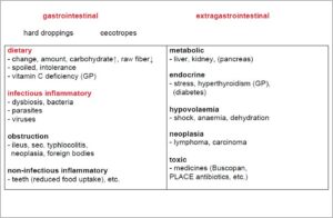
A complete medical history (duration, course, amount, admixtures) should always be taken at first, followed by a full clinical examination (weight, adspection, auscultation, palpation). In order to exclude gastrointestinal causes, a targeted faecal examination is the best diagnostic option and therefore the method of choice when working up a diarrhoeal disease. If extragastrointestinal causes (organic, metabolic-toxic, neoplastic) are suspected, a haematological blood test should be carried out, the clinical-chemical parameters should be determined and, if necessary, imaging techniques should be used (Fig.1).1
Faecal examination
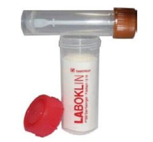
The collection of faecal samples is very simple. To facilitate the collection, use as little absorbent bedding as possible. For shipment to a laboratory, it is recommended to use suitable sample containers (1 sample tube per animal, approx. ¾ full) and to send them in an appropriate outer packaging to ensure hygienic shipment (Fig. 2).
Diagnostic options in the veterinary practice/fresh faeces
In the practice, you can already get a first indication of the cause of diarrhoea if you carry out a macroscopic faecal analysis (species-specific size and shape as well as colour, consistency and admixtures) and also examine the fresh (!) faecal sample microscopically directly on site.
- Fig. 1: Causes of diarrhoea in rabbits and guinea pigs (GP) (modified after Hein 2017)
- Fig. 2: Faeces tube with outer packaging
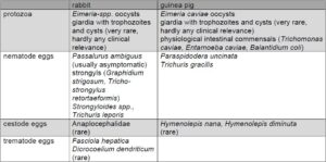
- Tab. 1: Endoparasites in rabbits and guinea pigs
- Tab. 2: Pathogenic bacteria in rabbits and guinea pigs
Fresh specimen
For a fresh specimen, a pea-sized amount of fresh faeces is usually sufficient. With – ideally warm – physiological saline solution or water, a faecal suspension is prepared. For the microscopic examination, a drop is placed on a slide and covered with a cover glass. Testing from a smaller sample quantity is generally possible, but carries the risk of reduced sensitivity.
Yeasts, such as Cyniclomyces guttulatus, as well as protozoa (Tab. 1) and occasionally worm eggs can be detected very well in the fresh specimen.
One of the most frequent causes of diarrhoea is dysbiosis – a shift in the rather gram-positive intestinal flora towards more gram-negative bacteria, clostridia and yeasts. Causes are often of dietary origin (inappropriate feeding, resulting in a lack of crude fibres and/or excess carbohydrates), any kind of malpositioned teeth or even parasitosis. A first and quick indication of a dysbiotic condition is the increased occurrence of yeast (>15/visual field) at 100-fold magnification.
Although flagellates, such as giardia, can be diagnosed in fresh faeces, they are very rarely found. Such findings in fresh faecal samples are only conclusive in positive cases. A more suitable test procedure for this is the giardia ELISA.
Tape test method
The tape test method – perianal tape impression using adhesive strips – is a quick and simple test method which may as well be carried out on site. It is particularly recommended for the detection of oxyurid eggs and larvae (Passalurus ambiguus). Detection by flotation is often negative, as oxyurid eggs tend to sink due to their high density. Sometimes, however, larval fragments of Passalurus ambiguus can be found in the flotation.
Pooled faeces
A 3-day pooled faecal sample generally increases the sensitivity of the faecal analysis for parasites (except for flagellates). When submitting faecal samples from rabbits or guinea pigs, different test methods are available: flotation, sedimentation (SAFC method), bacteriological examination, giardia ELISA and PCR (pathogen specific).
Flotation
The principle of the flotation test is based on the fact that parasite stages with a low density are transported to the surface of solutions which have a higher specific gravity. In rabbits and guinea pigs, this test method is suitable for the enrichment of coccidian oocysts as well as for the enrichment of species-specific nematode and cestode eggs (Tab. 1) and can be carried out in the practice, too, using commercially available test kits.
Sedimentation
During sedimentation, parasite stages with a high density settle in the sediment of a lighter solution. With this test method, especially trematode eggs (Fasciola hepatica, Dicrocoelium dendriticum) can be detected in rabbits. To some extent, the detection of nematode eggs is also possible, although flotation is to be preferred here due to its higher sensitivity. Trematode eggs have not yet been described in guinea pigs.
Bacteriological examination
The culture of faecal bacteria provides information on the pathogenesis and allows for targeted treatment through the creation of an antibiogram. Rabbits and guinea pigs as herbivorous caecal digesters physiologically have a mainly gram-positive flora with several hundred different bacterial species, yeasts, anaerobes and protozoa. Pathogen differentiation with a subsequent creation of an antibiogram is advisable for them, especially in food-producing animals and if human pathogenic organisms are suspected, but is of little use in the typical cases of diet-induced dysbiosis.
Shifts towards a gram-negative intestinal flora should always be considered in the context of feeding mistakes and/or parasitosis. This is because the detection of a certain species of bacteria is no proof that it is the actual cause of diarrhoea – except for pathogenic bacteria (particularly toxigenic ones) (Tab. 2).
Polymerase chain reaction
Polymerase chain reaction (PCR) is a method for the detection of specific pathogens, such as Lawsonia intracellularis. The advantage is its high sensitivity, the disadvantage is that it must be known beforehand which pathogen to look for and that only a positive result confirms it. A negative result cannot completely rule out an infection.
Blood test
Diarrhoea in rabbits and guinea pigs may also have extragastrointestinal causes. Depending on the animal species, various causes need to be considered, such as organic, metabolic or toxic disorders as well as hypovolaemia or neoplasia (Fig. 1). A blood test (Rodent Profile incl. a complete blood count) covers almost all differential diagnoses and should thus be part of the routine diagnosis of diarrhoea if the previous faecal examination was negative.
During the haematological examination of dysbiotic patients and patients with diarrhoea, a so-called pseudo left shift (shift from lymphocytic to neutrophil blood count) is often seen. Here, too, a differential blood count can help in making a diagnosis. In guinea pigs, leukocytosis with lymphocytosis indicates lymphoma and eosinophilia indicates parasitosis as a possible cause of diarrhoea. Unfortunately, this is not the case in rabbits; they usually have aleukaemic lymphoma and do not show eosinophilia due to parasites. A blood test is also suitable to clarify organic-metabolic or endocrine causes of diarrhoea (e.g. hyperthyroidism in guinea pigs). Hepatopathies (GLDH, AST, ALT, bilirubin, bile acids, triglycerides, cholesterol) and nephropathies (urea, creatinine) as a cause of diarrhoea as well as electrolyte shifts (Na, K, P), protein losses (total protein, albumin) and/or blood losses (erythrocyte count, haematocrit, total protein, albumin, urea) can easily be determined.
A blood test may also be very helpful in serious conditions such as ileus or obstruction: The glucose level serves as a prognostic factor in this case. The higher the level, the more likely it is that ileus is the cause and the worse the prognosis.2 If sodium is additionally considered in the interpretation, an even more precise prognosis can be made, because additional hyponatraemia doubles the mortality rate.3
Conlcusion
The faecal analysis combined with a haematological and clinical-chemical examination covers almost all the differential diagnoses of a diarrhoeal disease and can thus serve to initiate quick and targeted treatment.
Literatur:
-
1Hein J. (2017): Durchfallerkrankungen bei Kleinsäugern. Hannover, Schlütersche.
-
2Harcourt-Brown F M, Harcourt-Brown S F (2012): Clinical value of blood glucose measurement in pet rabbits. Vet Rec., 170(26):674.
-
3Bonvehi C et al. (2014): Prevalence and types of hyponatraemia, its relationship with hyperglycaemia and mortality in ill pet rabbits. Vet Rec., 174(22):554.


