Histopathological examinations of biopsies are an important tool for diagnosing gastrointestinal disease in dogs and cats.
The following will provide an overview of diseases of the gastrointestinal tract in which a histopathological examination of transmural or endoscopic biopsies can be helpful.
Anamnesis
Anamnestic information on the breed, age, clinical signs, feeding, previous treatment (e.g. cortison), results of diagnostic testing (e.g. ultrasound, parasitology, bacteriology, cobalamin and folic acid values, PLI, TLI) as well as findings during endoscopic sampling and tentative clinical diagnosis are important for the interpretation of the histopathological findings.
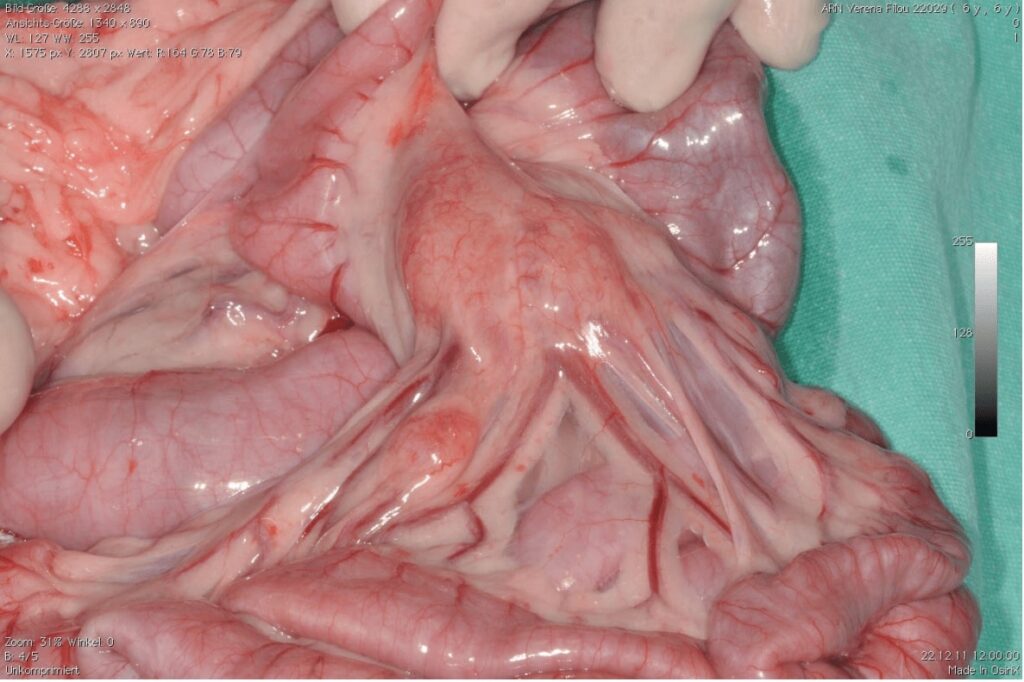
Transmural biopsies are taken during a laparotomy or laparoscopy. They have the advantage that they are not prone to loss of quality due to squeeze artifacts or suboptimal orientation of the sample and all of the layers of the intestinal wall can be evaluated. Laparotomy/laparoscopy also allows sample collection from additional sites (e.g. the jejunum), which are not accessible by endoscopy. It is also possible to sample other tissues such as liver, pancreas or lymph nodes (Fig. 1), or to aspirate the gall bladder.
Taking endoscopic biopsies is less invasive and allows an evaluation of the mucosal surface and targeted sampling of areas with lesions. It is best to take several samples that can be evaluated (up to 8 biopsies per localization).
The samples should be sorted according to the localization at which they were collected (ideally divided by plastic capsules on filter paper) and sent fixed in 4% formalin in suitable, labeled tubes.
Histopathological evaluation is carried out following processing of the samples using light microscopy on HE (hematoxylin and eosin) stained paraffin sections. The type and grade of mucosal lesions as well as inflammatory infiltration are evaluated according to the guidelines of the WSAVA (World Small Animal Veterinary Association Gastrointestinal Standardization Group; Day et al., 2008).
Helicobacter infections:
The pathological importance of Helicobacter spp. and so-called helicobacter-like organisms in dogs and cats is still debatable. Studies have shown that Helicobacter spp. can also be detected in healthy animals. There are controversial results on the influence of Helicobacter spp. on functional parameters in the stomach.
The pathogenic role of Helicobacter spp. in conjunction with a mild gastritis and clinically recurring vomitus is however, probable, especially when helicobacter are present in large numbers in biopsy material.
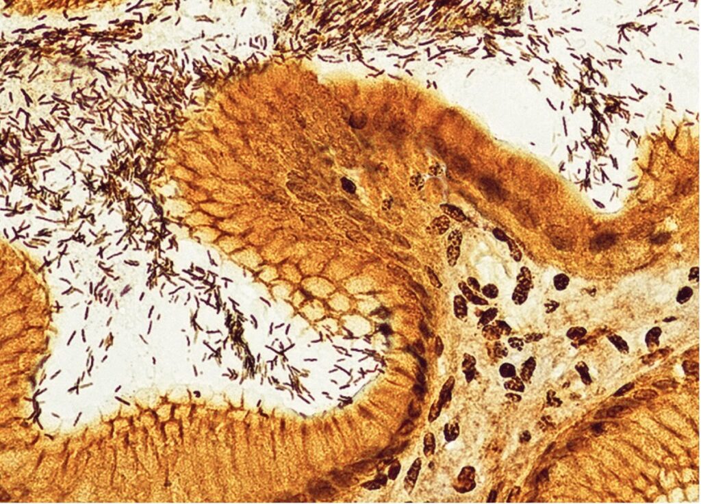
Noninvasive tests (antigen ELISA*) are available. In contrast, histopathological examination of biopsy material from the gastric mucosa has the advantage that the number of bacteria can be evaluated (Fig. 2). In addition, the possible presence of inflammatory changes can be reported. A gastritis (erosive ulcerative, mixed cellular, lymphoplasmacellular) indicating other causes can then also be evaluated.
It is always advisable to collect samples from multiple parts of the stomach (cardia, fundus, pylorus).
-
Fig. 1: Surgical situs with enlarged mesenterial lymph nodes; cat.
Picture Credits: Tierklinik Rupphübel, Beiwil am See, Switzerland
- Fig. 2: Helicobacter spp., Warthin-Starry stain, 400x
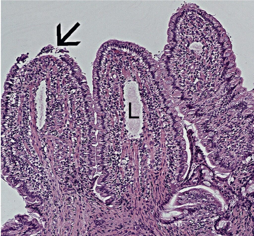
- Fig. 3: Histological lesions in a case of LPE (arrow points to epithelial lesion, L = mild lymphangiectases), HE 100x
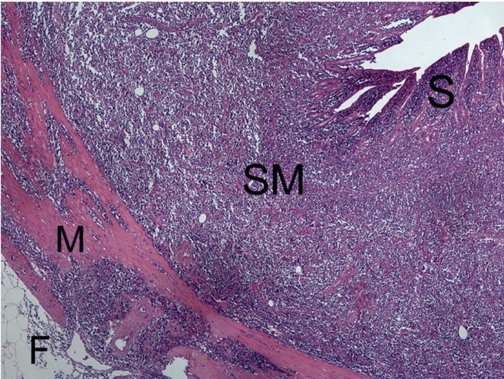
- Fig. 4: Intestinal lymphoma with transmural infiltration of tumor cells (S = mucosa, SM = submucosa, M = muscularis, F = fatty tissue)
Food incompatibility/ food allergy:
Food incompatibilities and allergies can cause gastrointestinal signs such as vomitius, diarrhea, and weight loss in dogs and cats.
In histological examinations, inflammatory infiltrations dominated by lymphocytes and plasma cells can be seen in the gastrointestinal mucosa. These cannot be differentiated from the lymphoplasmacytic form of inflammatory bowel disease (see below).
It is therefore always necessary to carry out an elimination diet (at least 4 to 6 weeks, commercially available elimination diets or homemade elimination food with one carbohydrate and one protein source) followed by a provocation diet in addition to a thorough food anamnesis. A serological allergy test* can be useful in choosing an appropriate dietary food.
Inflammatory bowel disease (IBD)
Inflammatory bowel disease is an idiopathic disease with persistent or recurring gastrointestinal signs in dogs and cats. Clinically, these can include vomiting, diarrhea, abdominal pain, inappetence, and weight loss.
Various forms of this disease have been described. These can affect different parts of the gastrointestinal tract and are also associated with different forms of inflammation.
Various factors that can lead to a loss of the immunological tolerance of the antigens in the intestinal lumen are the suspected cause of the disease (collapse of the mucosal barrier function, faulty regulation of the intestinal immune system, changes in the intestinal bacterial flora, genetic factors).
It is important to note that IBD is diagnosed by exclusion. The detection of histopathological changes in the intestinal mucosa is not diagnostic for IBD. Infectious and extraintestinal diseases as well as food incompatibilities (see above) must be ruled out before IBD can be diagnosed.
The following forms can be differentiated by histopathology:
- lymphoplasmacellular (gastro-) enteritis (LPE)
- lymphoplasmacellular colitis (LPC)
- eosinophilic gastroenteritis (EGE)
- granulomatous colitis
LPE/LPC are the most common forms of IBD. Histopathologically, these are characterized by infiltration of lymphocytes and plasma cells, shortening and fusion of the villi, epithelial lesions, and crypt lesions as well as a moderate lymphangiectasia (Fig. 3).
EGE is the second most common form of IBD. Inflammatory infiltrates of the mucosa dominated by eosinophilic granulocytes, atrophy and fusion of the villi are characteristic.
Granulomatous forms of IBD are rare.
Histiocytic ulcerative colitis (HUC) is found almost exclusively in boxers and is no longer included in IBD, since an association with invasive Escherichia coli has been proven.
Eosinophilic enteritis / hypereosinophelia syndrome
An eosinophilic gastroenteritis can occur as a form of IBD (see above), parasitological, or allergic (food allergy).
A systemic disease in the form of a hypereosinophilia syndrome is also observed in cats and more rarely in dogs.
A hypereosinophilia syndrome is associated with eosinophilic infiltrations in multiple tissues as well as a blood eosinophilia.
In many cases, the gastrointestinal tract is also affected and the animal develops clinical gastrointestinal signs. A hypertrophy of the muscularis can often be detected (sonographically).
Histopathologically, well-differentiated eosinophilic granulocytes are visible throughout the intestinal wall. If hypereosinophilia syndrome is suspected, it can be helpful to also examine samples from the liver and spleen.
Protein losing enteropathy (PLE)
Inflammatory intestinal diseases that are clinically associated with a severe loss of protein are found most commonly in Shar-Peis, German Shepherds, Rottweilers, and Yorkshire Terriers.
A disease that is probably hereditary has been described in Soft Coated Wheaten Terriers and manifests as a protein losing enteropathy and/or protein losing nephropathy (PLN). A genetic test* can be carried out for PLN in this breed.
Severely dilated lymph vessels in the intestinal villi and in deeper layers of the intestinal wall as well as infiltration of foamy macrophages are typical histopathological changes in PLE. Lipogranulomas can sometimes be found in the muscularis. Macroscopically, these appear as small white growths on the serosa and in the mesenterium.
Differential diagnoses include bacterial gastroenteritis, intestinal mycoses, neoplasias, and chronic foreign body reactions which can be associated with a protein losing enteropathy. In cats, protein losing enteropathies are less common and are often associated with intestinal lymphomas.
Neoplasia:
Clinically, palpation and imaging (ultrasound), among others, can lead to a suspected diagnosis of gastrointestinal neoplasia. Histopathology is necessary in order to confirm the diagnosis (rule out inflammation, e.g. caused by a foreign body), as well as to determine the type and behavior of the neoplasia.
Tumors of the gastrointestinal tract are mostly intestinal lymphomas, adenocarcinomas, leiomyomas, leiomyosarcomas, and gastrointestinal stromal tumors (GIST).
If nodular neoplasias are completely resected, the excisional margines can be histologically examined. For biopsies, it is important to remember that neoplasias are often associated with severe ulcerative-inflammatory lesions and infiltrative growth (esp. carcinomas of the stomach and rectum) may only be reliably histopathologically detectable in deep tissues.
Intestinal lymphomas are the most common neoplasias of the gastrointestinal tract in dogs and cats. Intestinal lymphomas can affect deep layers of the intestinal wall (Fig. 4). Transmural biopsies are preferable for diagnostics, since early stages may be difficult to differentiate from LPE in the mucosa.
Differentiation of the cellular origin (T or B cell lymphomas) can only be carried out by immunohistological examination or by determining the lymphocytic clonality using PARR (PCR for antigen receptor rearrangement).
Rectal polyps/carcinomas are found mostly in middle aged and old dogs. Adenomatous polyps can develop into in situ carcinomas and highly malignant carcinomas. Superficial biopsies are often non diagnostic in such cases, since invasive growth in the submucosa and muscularis are significant diagnostic and prognostic characteristics.
Check list for gastrointestinal biopsies:
Anamnesis
- Age, breed
- Clinical signs
- Findings of other diagnostic tests
- Suspected diagnosis
- Previous treatments (esp. with cortison)
Sampling
- Representative location
- Sufficient number of biopsies
- Gentle treatment of biopsy material
- Submit/fix biopsies sorted according to collection site in labeled tubes
- Fix samples in 4% formalin
*Testing can be carried out by Laboklin


