










Introduction
Anaemias are among the most common haematological abnormalities encountered in small animal medicine. While regenerative anaemias can generally be attributed relatively clearly to blood loss or haemolysis, non-regenerative anaemias often present a diagnostic challenge. Interpretation becomes particularly complex when alterations in iron metabolism are present. Iron deficiency can result from chronic blood loss, but can also be a manifestation of inflammatory or chronic disease. Distinguishing between absolute and functional iron deficiency is essential for treatment. As the underlying pathophysiological mechanisms differ, they have a significant impact on both diagnostic approaches and therapeutic decisions.
The classical serum iron value alone has very limited diagnostic significance. Both patients with true iron deficiency resulting from chronic blood loss and animals with anaemia of chronic disease may have low serum iron concentrations. Only the combined interpretation of additional parameters of iron metabolism allows for a more differentiated assessment.
-
-
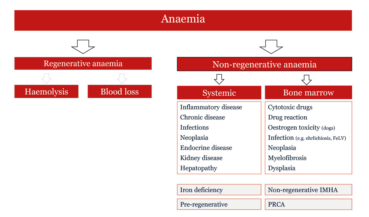
Fig. 1: Differential diagnoses for anaemia
Image source: Dr Jennifer von Luckner
Non-regenerative Anaemia: What Next?
The classification of anaemia begins with the question of whether regeneration, or effective erythropoiesis, is present. Increased absolute reticulocyte counts indicate a regenerative anaemia and therefore suggest blood loss or haemolysis as the underlying cause. If a reduced haemoglobin concentration combined with a low haematocrit is not accompanied by reticulocytosis (= ineffective erythropoiesis), a non-regenerative anaemia is present.
Non-regenerative anaemias have a broad differential diagnosis (Fig. 1). These include:
- inflammatory diseases,
- chronic diseases,
- infections,
- neoplasia,
- endocrinopathies,
- chronic kidney disease,
- hepatopathies,
- bone marrow disorders,
- drug-induced changes,
- iron
Caution: Not all anaemias that initially appear to be non-regenerative actually belong to the classic category of non-regenerative anaemias. This is particularly relevant in two situations:
-
- pre-regenerative anaemias following acute blood loss or haemolysis,
- chronic bleeding anaemia with progressive iron
Following acute blood loss, the bone marrow requires approximately three to five days before a measurable reticulocytosis develops. An initially non-regenerative anaemia therefore does not rule out blood loss or haemolysis.
In chronic blood loss, on the other hand, progressive iron loss occurs. Initially, the anaemia is still regenerative; later, regeneration becomes inadequate and is eventually completely suppressed because the bone marrow no longer has sufficient iron available for haemoglobin synthesis.
Iron Metabolism
Iron plays a central role in the body, not only in erythropoiesis. The body therefore tightly regulates the absorption, transport, storage and release of this trace element.
Intestinal iron absorption takes place predominantly in the duodenum and proximal jejunum. Haem iron from animal sources is absorbed more efficiently than non-haem iron from plant sources. Following absorption, iron is released from enterocytes via ferroportin and transported bound to transferrin.
Transferrin acts as a transport protein, delivering iron to tissues with a high demand for iron, particularly the bone marrow. Iron is stored primarily in the form of ferritin in macrophages and hepatocytes. Ferritin is the body’s main iron storage protein.
The principal regulator of iron metabolism is hepcidin, a peptide hormone produced in the liver. Hepcidin inhibits ferroportin, thereby reducing intestinal iron absorption in particular. It also blocks the release of iron from macrophages and the mobilisation of stored iron.
Inflammatory processes, chronic diseases or neoplasia often lead to increased hepcidin production and consequently reduced iron availability despite adequate iron stores.
Absolute versus Functional Iron Deficiency
Absolute Iron Deficiency
In absolute iron deficiency, the body’s iron stores are genuinely “empty”. Causes include increased iron loss or utilisation (acute or chronic blood loss), increased iron requirements (e.g. during pregnancy), insufficient dietary iron intake (inadequately supplemented vegetarian diets) or impaired absorption (e.g. severe chronic intestinal disease).
The most commonly described cause in dogs is chronic blood loss. From a pathophysiological perspective, this results in a true loss of iron from the body.
Typical causes include:
- chronic gastrointestinal bleeding,
- bleeding tumours,
- ulcers,
- parasitic infections,
- chronic haematuria,
- thrombocytopenia (other coagulopathies less commonly result in chronic blood loss).
Functional Iron Deficiency
In functional iron deficiency, the body has sufficient iron available, but does not make it available to the bone marrow. This form typically occurs in chronic inflammation, infections, neoplasia and severe systemic diseases. The underlying hepcidin-mediated iron retention means that iron remains stored in macrophages and storage sites.
Diagnostics and the Iron Profile
Iron deficiency leads to inefficient haemoglobin synthesis in the bone marrow. The erythrocytes are smaller and contain less haemoglobin. In addition, erythropoietic activity is reduced (increased apoptosis and increased oxidative stress on the cells).
The initially purely regenerative anaemia caused by blood loss gradually develops into a non-regenerative anaemia due to insufficient erythrocyte production. This transition occurs gradually, progressing from a regenerative to an inadequately regenerative and finally to a non-regenerative anaemia.
As this process progresses, further signs of iron deficiency may be observed:
-
- on the blood smear, hypochromic (pale erythrocytes with low haemoglobin content) and microcytic (small) erythrocytes may be observed;
- in the blood count:
-
- changes in erythrocyte indices:
- MCH (mean corpuscular haemoglobin) decreased,
- MCHC (mean corpuscular haemoglobin concentration) decreased,
- MCV (mean corpuscular volume) decreased,
- changes in erythrocyte indices:
- reticulocyte haemoglobin (Ret-He/Ret-Hb/ ReticHb) decreased,
- serum iron concentration decreased,
- hypoproteinaemia (in cases of blood loss, both albumin and globulin are often decreased),
- an isolated increase in urea may occur with intestinal blood loss (postprandial elevation must be ruled out).
-
The signs of functional iron deficiency can be very similar. This can make it difficult to differentiate between absolute (= blood loss) and functional (= inflammation/chronic disease) iron deficiency.
As a result, there may be uncertainty regarding the appropriate further diagnostic approach and therapeutic consequences.
In human medicine, several parameters of iron metabolism are used to address this question. These parameters are now also available for veterinary medicine, at least for dogs (Table 1).
Hepcidin
Hepcidin leads to reduced intestinal iron absorption and iron retention in the reticuloendothelial system. As expected, it is increased in functional iron deficiency. Conversely, low hepcidin levels promote iron absorption and mobilisation of stored iron. Hepcidin is therefore low in absolute iron deficiency.
Ferritin
Ferritin is a protein complex that serves as an iron store. It is also often referred to as “storage iron” or an “iron depot”. In absolute (“true”) iron deficiency, the body’s iron stores are depleted and the ferritin concentration in the blood is low. In functional iron deficiency, on the other hand, the concentration will be normal or even increased.
Unsaturated Iron-Binding Capacity (UIBC)
UIBC measures the amount of iron that can still bind to the available transferrin. The more iron that can still be bound, the less iron was previously present, meaning that the transport protein transferrin is relatively “empty”. In absolute (“true”) iron deficiency, an increased UIBC value is therefore expected.
Total Iron-Binding Capacity (TIBC)
This value is calculated and represents the total amount of iron that can bind to the available transferrin. TIBC correlates with the concentration of transferrin in the blood. The higher the TIBC, the more transferrin is present. Transferrin concentrations in the blood are often decreased as part of a negative acute-phase response in inflammation and tumours, but may also be decreased in gastrointestinal absorption disorders. Therefore, in anaemia of chronic disease/ inflammatory disease associated with functional iron deficiency, TIBC is expected to be reduced.
Table 1: Iron profile parameters and their changes in absolute and functional iron deficiency
| Parameter | True iron deficiency (blood loss | Functional iron deficiency (inflammation, tumour, infection) |
| Eisen | ⇓ | ⇓ |
| Ferritin | ⇓ | ⇑ / = |
| Hepcidin | ⇓ | ⇑ |
| UIBC (unsaturated iron binding capacity) |
⇑ | ⇓ / = |
| TIBC (total iron binding capacity) |
⇑ | ⇓ / = |
| Iron saturation | ⇓ | ⇑ / = |
Iron Saturation
Iron saturation is also calculated. It is also referred to as transferrin saturation and is a measure of the amount of iron bound to circulating transferrin. The higher the value, the more iron is transported by transferrin and the more iron is available in the body. In absolute iron deficiency, low iron saturation is expected, whereas in functional iron deficiency, it should be normal or even increased.
Conclusion: There is no single perfect parameter for diagnosing iron deficiency. Rather, several parameters must always be considered together and interpreted in the context of the clinical question. In addition to the blood count, the iron profile can help differentiate between absolute (true) and functional iron deficiency. This is particularly important when investigating the underlying cause and selecting the appropriate treatment.
Jennifer von Luckner, Annemarie Baur-Kaufhold
Our Diagnostic Services for Iron Deficiency:
- Complete blood count
- Cytological blood smear examination
- Basic anaemia screening, dogs and cats
- Iron metabolism profile
Further reading:
-
Mead MK, Claus M, Litton E, Smart L, Raisis A, Rossi G, Trengove RD, Gummer JPA. Identification of the canida iron regulatory hormone hepcidin. Sci Rep. 2019; 18 (9): 19400.
-
Pfeiffer C, Looker AC. Laboratory methodologies for indicators of iron status: strengths, limitations, and analytical challenges. Am J Clin Nutr. 2017; 106, Suppl 6: 1606S-1614S.
-
Ramos CF, Doulidis PG, Polakova N, Burgener IA, Jensen-Jarolim E, Cimarelli G, Panakova L, Roth-Walter F. Iron deficiency in dogs suffering from atopic dermatitis. BMC Vet Res. 2024; 20 (1): 506.
-
Rohr M, Brandenburg V, Brunner-La Rocca H-P. How to diagnose iron deficiency in chronic disease: A review of current methods and potential marker for the outcome. Eur J Med Res. 2023; 28: 15.
-
Rusch JA, van der Westhuizen DJ, Gill RS, Louw VJ. Diagnosing iron deficiency: Controversies and novel metrics. Best Pract Res Clin Anaesth. 2023; 37 (4): 451-467.
-
Vizi Z, Lanyi K, Bagi M, Laczay P, Balogh N, Sterczer A. Serum hepcidin measurements in healthy dogs using liquid chromatography/tandem mass spectrometry. Vet Clin Pathol. 2020; 49 (2): 292-298.