In dogs and cats, increased urinary protein excretion is pathological and highly correlated with reduced survival of the respective animal. The excretion of small quantities of albumin < 1 mg/dl is considered physiological in dogs and cats.
A urine protein-creatinine ratio (UP/C) of > 0.4 in cats and > 0.5 in dogs is defined as proteinuria according to the guidelines of the IRIS (International Renal Interest Society). A distinction is made between prerenal, renal and postrenal proteinuria. Their various causes are shown in Table 1.
Urine protein electrophoresis has proven to be a useful tool to differentiate proteinuria. Here, the diluted urine is placed on a gel, the proteins are separated according to molecular weight and it is then possible to distinguish the type of proteinuria from the protein pattern.
Prerenal proteinuria
The excretion of light-chain antibody fragments, so-called Bence Jones proteins, indicates highly pathological prerenal proteinuria. They are produced if multiple myeloma is present and usually cause high-grade proteinuria.
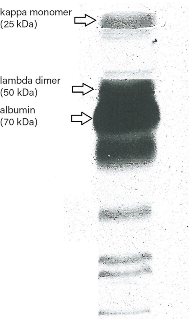
In urine electrophoresis, the patterns of kappa monomers (25 kDa) and lambda dimers (50 kDa) are depicted (Fig. 1). Other common causes of prerenal proteinuria are hypertension, hyperadrenocorticism in dogs and hyperthyroidism in cats.
Tab. 1: Proteinuria: possible causes and diseases (according to: Harley and Langston, 2012)
| cat | cat & dog | dog | |
| prerenal | hyperthyroidism | multiple myeloma systemic hypertension drug reactions acute pancreatitis |
hyperadrenocorticism |
| renal | hyperthyroidism
any severe inflammatory neoplasia, infectious and immune-mediated disease |
acute renal failure chronic kidney disease glomerulopathy acute pancreatitis viral disease drug reactions systemic hypertension Diabetes mellitus endocarditis |
hyperadrenocorticism
immune-mediated disease (systemic lupus erythematosus, immune-mediated haemolytic anaemia, |
| postrenal | lower urinary tract disease reproductive tract disease |
Renal proteinuria
For one, this includes glomerulopathies in which high molecular weight proteins larger than albumin (> 70 kDa) can no longer be retained by the glomerular apparatus due to various defects.
Glomerulopathies are associated with a variety of infectious (e. g. ehrlichiosis, babesiosis and leishmaniasis in dogs; FIV, FELV and FIP in cats), neoplastic, parasitic, autoimmune and endocrine causes.
Furthermore, we distinguish tubulopathies in which the capacity or ability of the proximal tubule to reabsorb low molecular weight proteins (< 70 kDa) is exhausted.
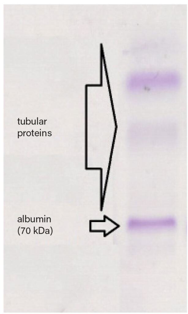
These tubulopathies manifest as Fanconi syndrome in dogs, which is hereditary in Basenjis and caused by toxic factors in other breeds, e. g. after the administration of gentamicin (Fig. 2).
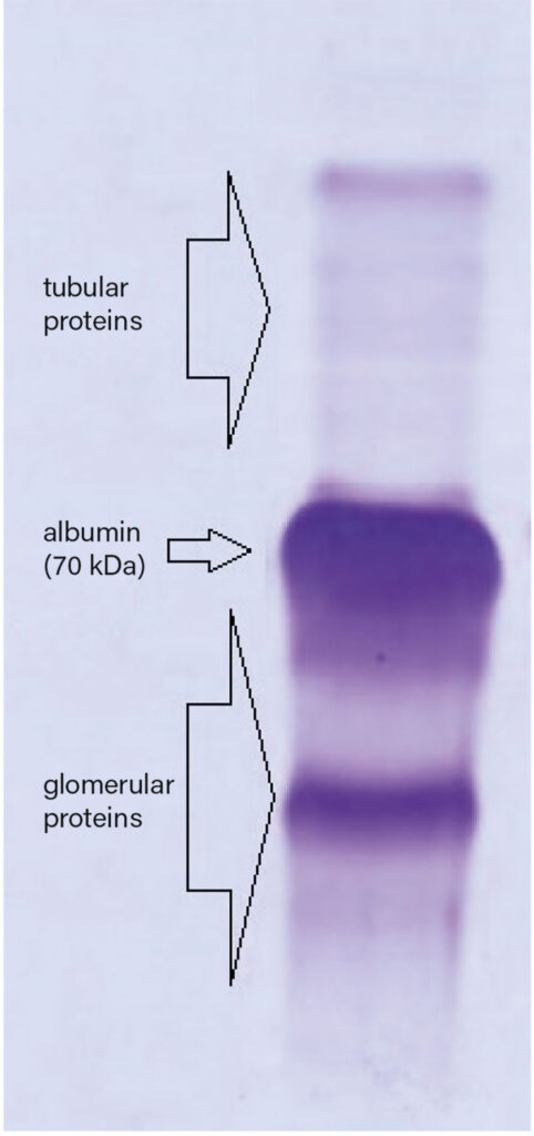
Mixed proteinuria, in which both protein groups can be detected, mainly occurs in stages 2 – 4 of chronic kidney disease and in acute kidney injury because, in addition to glomerular dysfunction, it also causes renal tubular acidosis (Fig. 3).
Since treatment of proteinuria can vary greatly depending on the cause, it is essential to determine whether renal proteinuria is glomerular, tubular or mixed glomerular-tubular proteinuria.
Postrenal proteinuria
This is usually caused by lower urinary tract and reproductive tract disease, can be detected by examination of urine sediment and does not require differentiation by urine electrophoresis.
-
Fig. 1: Urine electrophoresis of a cat (7 years) with multiple myeloma: The patterns of kappa monomers and lambda dimers can be seen. Due to the advanced renal damage, albumin and glomerular proteins can also be detected.
Photo credits: Laboklin
-
Fig. 2: Urine electrophoresis of a Miniature Bull Terrier (6 months) with Fanconi syndrome caused by toxicity: Albumin and tubular proteins only can be detected.
Photo credits: Laboklin
-
Fig. 3: Urine electrophoresis of a cat (16 years) with chronic kidney disease: Both tubular and glomerular proteins can be detected in the urine.
Photo credits: Laboklin
Urine electrophoresis at Laboklin
Thanks to an updated methodology, the excreted proteins can be presented in a more differentiated way. We now offer you an evaluation of the urinary proteins in which all relevant glomerular and tubular proteins are differentiated and interpreted, so that urine electrophoresis can provide even more specific information about the underlying disease.
For this test, we need 1 ml of urine; the test duration is 3 – 5 days. The examination is useful if the UP/C ratio is increased.
As always, our experts from the veterinary team are available by telephone to help you with the interpretation.
Dr. med. vet. Marco Weiß
References
-
Harley L, Langston C. Proteinuria in dogs and cats. Can Vet J. 2012; 53: 631–638.
-
Vaden SL. Glomerular disease. In: Ettinger SJ, Feldman EC. Textbook of Veterinary Internal Medicine. 6th ed. St. Louis, Missouri: Saunders (Elsevier). 2005:1786–1800.
-
International Renal Interest Society (IRIS) Staging of CKD. modified 2019.
-
Brown SA. Renal tubular Defects in Small Animals. MSD Veterinary Manual 2016.


