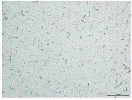
Dental disease and inappropriate feeding are common causes of diarrhoea. Parasites, in contrast, play a smaller role in pets. However, some parasites may occur more commonly depending on the type of husbandry and age of the animals (larger collections, breeders, outdoor enclosures, juvenile animals). While young animals often develop clinical signs when they are infested, adults may only develop notable changes in cases of severe parasitism. Parasite infestation can, however, lead to changes and disruptions in the intestinal environment. Increased growth of yeasts (Fig. 1) or bacterial secondary infections (e.g. with Clostridia, E. coli) can be a result and can cause intestinal disease.
Parasites of rabbits
Protozoa
Coccidia
Various Eimeria species can infest the intestine or bile ducts of rabbits. Young rabbits are particularly susceptible to intestinal coccidiosis, and infestation in these animals can be associated with high mortality. Infestations can spread epidemically within collections. Adult animals can be clinically inapparent shedders. In addition to diarrhoea, animals can develop bloat and appear dull and lose their appetite. In bile duct coccidiosis, severe infestation leads to a reduction in liver function. Affected animals are apathetic, lose weight, and are constipated. Some animals may also develop fever and become icteric.
Treatment
Treatment of coccidiosis is carried out with toltrazuril (10 mg/kg once daily per os for 3 days, repeat after 3 days). Sulfonamides such as sulfadimethoxine can also be used, but are often less effective than toltrazuril.
Giardia
Giardia infections are rare in rabbits. The occasional diarrhoea is usually slimy and has a light colouration. Detection via SAF is preferable to the flotation method. Coproantigen ELISAs are even more sensitive.
Treatment
Fenbendazole (20 mg/kg once daily per os) or metronidazole (10-20 mg/kg twice daily per os) can be used in infected rabbits for at least 5 days.
Nematodes
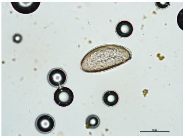
Of the nematodes, Passalurus ambiguus (pinworm) is most common (Fig. 2). Animals often only develop clinical signs (diarrhoea, bloat, colicky abdominal pain, anal itching) if they are severely parasitized. Normal faecal exams can be false negative. In suspected cases it is therefore prudent to prepare a cellophane tape impression of the anus, since eggs are laid on the rectal and anal mucosa and on the surface of faecal pellets.
Stronglyus-type eggs can also be found in the faeces, and can be from Graphidium strigosum or Trichostrongylus retortaeformis. Like Strongyloides spp. and Trichuris leporis, these parasites play a subordinate role in pets. Animals can be infected by feeding greens that have been contaminated by wild rabbits or field hares, or by the use of outdoor enclosures. Young animals are mostly affected with diarrhoea, dullness, and inappentence.
Treatment
Animals with nematode infestations can be treated with (pro-) benzimidazoles like fenbendazole (20 mg/kg once daily per os for 5 days, repeat after 14 days), mebendazole (20 mg/kg once daily per os for 3-5 days, repeat after 14 days), or febantel (10 mg/kg once daily per os for 3 days, repeat after 14 days). Subcutaneous administration of ivermectin (0.3-0.5 mg/kg) or doramectin (0.5 mg/kg) repeating after 7-14 days is also possible.
Cestodes
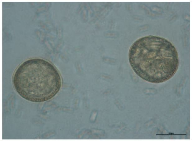
Tapeworm infestations are rare in wild rabbits, and even rarer in pet rabbits. Tapeworms in the family Anoplocephalidae (Fig. 3) are transmitted by oribatida (moss or beetle mites), which are intermediate hosts. Clinical signs are found mostly in juveniles or in cases of mass infestations.
Treatment
Praziquantel (one treatment with 10 mg/kg per os or subcutaneously, repeat after 10-14 days) can be used to treat tapeworm infestations.
- Fig. 1: Cyniclomyces guttulatus
- Fig. 2: Passalurus ambiguus
- Fig. 3: Tapeworm egg from the family Anoplocephalidae
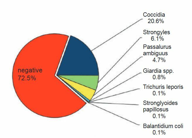
- Fig. 4: Parasite detection in rabbits using flotation and SAF methods (n=3746)
- Fig. 5: Paraspidodera uncinata
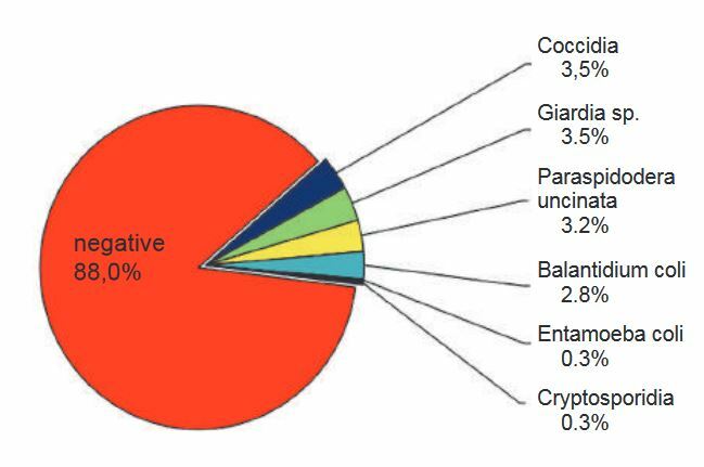
- Fig. 6: Parasite detection in guinea pigs using flotation and SAF methods (n=689)
Trematodes
Infestations with Fasciola hepatica or Dicrocoelium dendriticum are extremely rare. Transmission is via green fodder or swampy locations or sheep pastures, whereby the metacercariae of Fasciola hepatica adhere directly to the fodder, while the metacercariae of Dicrocoelicum dendriticum are ingested through infested ants. The common liver fluke can cause hepatitis and cholangitis, leading to inappetence, wasting, icterus, and oedema formation. Infestations with the lancet liver fluke generally remain undetected.
Treatment
Treatment with substances such as e.g. closantel (10 mg/kg per os, single dose) for Fasciola hepatica or fenbendazole (100 mg(kg per os) for Dicrocoelium dendriticum have been described.
Incidence of parasites in rabbits
An evaluation of routine submissions (n=3746) showed that parasites were detected in 27.5% of the rabbits, 72.5% of the rabbit samples were negative (Fig. 4).
Parasites of guinea pigs
Protozoa
Trichomonads
Trichomanads are physiological intestinal commensals in the caecum and colon of healthy guinea pigs. They can, however, strongly proliferate and cause disease if the intestinal environment changes or in immunosuppressed animals. Changes in the intestinal environment can be caused e.g. by other parasites, incorrect feeding or dental disease. The resulting chronic diarrhoea is soft and the animals lose weight. Trichomonads can be detected in native smears of fresh faecal material. It is important to determine the cause of the proliferation of flagellates.
Treatment
Metronidazole and dimetridazole (20-50 mg/kg once or twice daily per os) can be used for 7 days to treat trichomonads.
Entamoeba caviae and Balantidium coli
These single celled organisms are also commensal in the caecum and colon. As for trichomonads, they can proliferate and cause disease under various circumstances. An increased detection of Balantidium coli, for example, is an indication of too little structure in the fodder.
Treatment
Metronidazole and dimetridazole can also be used to treat these flagellates.
Cryptosporidia
Cryptospoidium wrairi plays a minor role in pets. Increased rates of infestation have been described in large collections. In suspected cases, a coproantigen ELISA is better for diagnosis than flotation.
Treatment
There are no effective treatment.
Giardia
Giardia are rare in guinea pigs. Affected animals generally do not have diarrhoea. SAF is superior to flotation for detection. Coproantigen ELISAs are even more sensitive.
Treatment
Fenbendazole (20 mg/kg once daily per os) or metronidazole (20-40 mg/kg twice daily per os) for 5 days can be used to treat infected guinea pigs.
Coccidia
Infestation with Eimeria cavia is mostly relevant in groups, such as breeding groups or in the animal trade. Juveniles most commonly develop disease. Affected animals are apathetic, inappetent, lose weight, and have diarrhoea, with high mortality in some cases.
Treatment
The same treatment is used in guinea pigs as in rabbits.
Nematodes
Infestation with Paraspidodera uncinata (pinworm) is mostly found in large collections, outdoor enclosures or in animals with outdoor runs. Clinical signs manifest in cases with severe infestations (Fig. 5).
Trichuris gracilis is found mostly in wild guinea pigs, but has also been described in pet animals in individual cases.
Treatment
Various substances are effective against nematodes. Of the (pro-)benzimidazoles, e.g. fenbendazole (20 mg/kg once daily per os for 5 days, repeat after 14 days), and febantel (10 mg/kg once daily per os for 3 days, repeat after 14 days) can be used. Subcutaneous injection of ivermectin (0.3-0.5 mg/kg) or doramectin (0.5 mg/kg) with a single repeat treatment after 7-14 days are also effective.
Cestodes
Hymenolepis nana or Hymenolepis diminuta can be found in guinea pigs, but are quite rare. Insects are the intermediate hosts. Transmission to guinea pigs is via oral ingestion of these insects (e.g. fleas, flour beetles, mealworms, cockroaches). Hymenolepis nana can also be transmitted by direct oral ingestion of eggs. Infestation is usually clinically inapparent.
Treatment
Infestation is treated with praziquantel (single administration of 5-10 mg/kg per os or subcutaneously, repeat after 10-14 days).
Incidence of parasites in guinea pigs
An evaluation of routine submissions (n=689) showed that parasites were detected in 12.0% of the guinea pigs, 88.0% of the samples were negative (Fig. 6).
