Turtles and tortoises are popular pets which can become very old when kept properly. Apart from the commonly kept European species, such as Hermann’s tortoise (Testudo hermanni) (Fig. 1), experienced owners also enjoy keeping exotic species like radiated tortoises (Astrochelys radiata) or leopard tortoises (Stigmochelys pardalis). Clinical signs often appear very late and are only very non-specific in chelonians. Thus, in addition to the standard clinical examination and imaging, laboratory testing is the most important diagnostic tool for providing an accurate diagnosis quickly and at an early stage. This is particularly important in late summer for animals from temperate zones, as a detailed diagnosis is recommended before brumation so that a “rude awakening” or no awakening at all during and after brumation can possibly be avoided. The most helpful laboratory tests include haematology and biochemistry, parasitology, molecular biology and serology.
Blood testing in chelonians
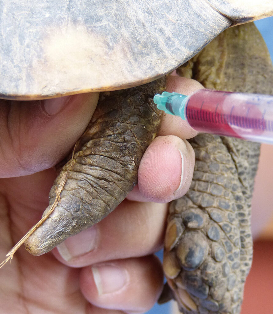
Lithium heparin blood is best suited for blood testing in chelonians (Fig. 2). Clinical chemistry is particularly important for monitoring organ function, as hepatic or renal impairments can be dangerous for the animals, especially during brumation.
Depending on the species, gender, season and temperature, there may be changes in the individual clinical chemistry parameters that need to be taken into account. Contamination with lymph causes reduced protein levels, liverassociated enzymes, uric acid levels and a shift in electrolytes. Trauma during blood collection, for example after several unsuccessful venipuncture attempts, can lead to an increase in alkaline phosphatase and creatine kinase. Considering the wide range of normal values in some species, it is advisable that blood testing is also carried out on healthy animals, so that it is easier to compare and interpret the results if the animal is ill.
Kidney diseases
Uric acid, the major end product of protein and purine metabolism in terrestrial species, is the main indicator of kidney disease. There are strong increases in various kidney diseases, gout, renal dysfunction due to bacteraemia and septicaemia as well as in kidney necrosis due to nephrotoxic drugs such as aminoglycosides and sulphonamides. Feeding animal-based food, as it is physiological in some aquatic turtle species, can also lead to an increase in uric acid. Lowered levels are associated with hepatic diseases. Urea only plays a minor role in terrestrial species, as the levels remain within the normal range for a long time even if uric acid levels are high. In aquatic species, urea also is an excretory product of protein metabolism, which is why it plays a greater role in the diagnosis of kidney diseases. In kidney diseases, blood phosphate levels increase as well, but hyperphosphataemia can also be caused by excessive dietary intake, hypervitaminosis D and haemolysis. In young animals, phosphate levels as well as calcium and alkaline phosphatase are physiologically increased as a result of bone growth.
Hepatic diseases
GLDH (glutamate dehydrogenase) is the main indicator of liver cell damage. It naturally occurs in the mitochondria of liver cells and is only released into the blood when the cells are destroyed. Other enzymes also found in liver cells are ALT (alanine amino transferase), AP (alkaline phosphatase) and AST (aspartate amino transferase). However, these enzymes are not only found in the liver, but also in other organs of the body, so they are not very specific and have to be interpreted in combination with other blood test results and physiological conditions. Liver function parameters are substances that are naturally synthesised by the liver and are decreased or increased if the liver function is disturbed. In this context, especially bile acid should be mentioned, though it can also be affected by feeding, bile duct obstruction and dehydration. Other functional parameters are the protein fractions in the blood, which are, however, also influenced by food intake and renal function. For diagnosing steatosis, it may be useful to determine the triglyceride and cholesterol levels in the blood, as these are usually elevated in this context. However, it should be noted that they are physiologically elevated in females during yolk formation (vitellogenesis).
Haematology
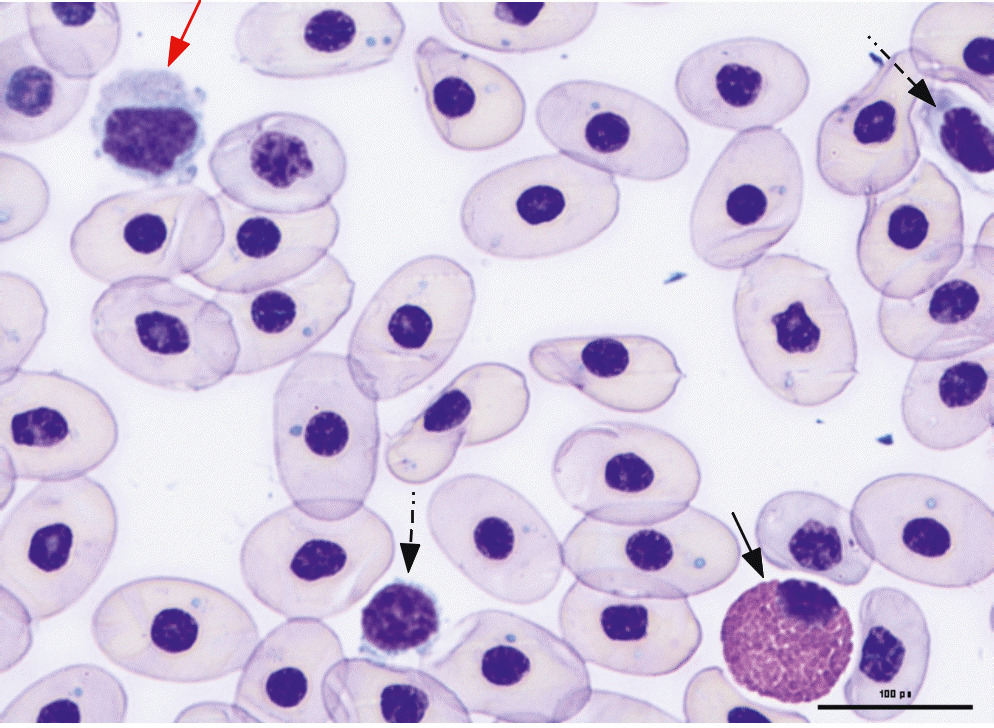
By determining the level of haematocrit, anaemia or dehydration can be diagnosed. A haematological analysis can also provide important information on inflammation. Shifts in cell count are less pronounced in reptiles than in mammals and are therefore more difficult to interpret. However, bacterial and parasitic infections as well as stress can lead to an increase in heterophilic granulocytes, too. Eosinophilic granulocytes are increased in parasitic infections and when the immune system is stimulated. An increase in lymphocyte count occurs during wound healing, inflammation as well as parasitic and viral infections. Changes in the morphology of individual cells can also indicate infections. When looking at the smear, parasites or inclusions due to viral or bacterial infections can be diagnosed as well, but they should not be confused with artefacts caused, for example, during the preparation or drying of the smear. Species, gender, age and physiological condition of the animal also influence the total count and the ratio of the different leucocytes. It is, however, also important to note that physiologically there can be strong seasonal fluctuations in cell distribution.
-
Fig. 1: Hermann’s tortoise (Testudo hermanni)
Photo credits: PD Dr. Rachel E. Marschang
-
Fig. 2: Blood collection from the dosal tail vein of a marginated tortoise (Testudo marginata)
Photo credits: PD Dr. Rachel E. Marschang
-
Fig. 3: Blood smear of a red-eared slider (Trachemys scripta elegans). A lymphocyte (red arrow), an eosinophilic granulocyte (black arrow) as well as two platelets (black, dashed arrow) and some staining artefacts in the erythrocytes can be seen.
Photo credits: Laboklin
-
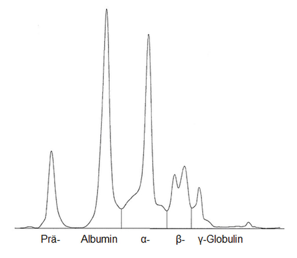
Fig. 4: Capillary electrophoresis analysis of plasma of a healthy Hermann’s tortoise (Testudo hermanni)
Photo credits: Laboklin
-
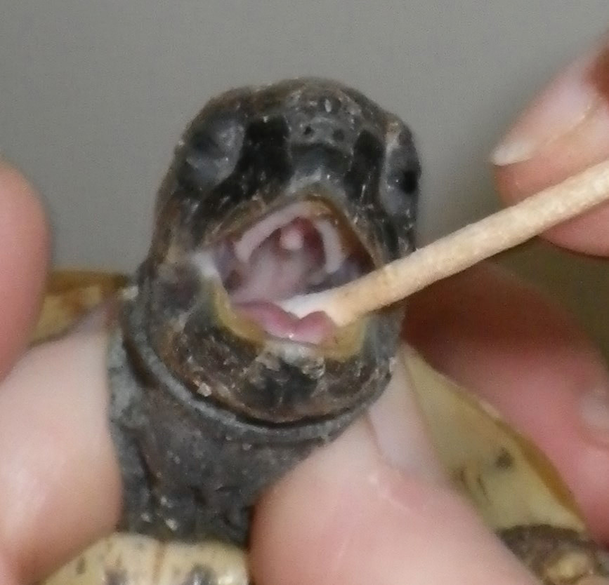
Fig. 5: Pharyngeal swab sampling in a Hermann’s tortoise (Testudo hermanni)
Photo credits: PD Dr. Rachel E. Marschang
Electrophoresis
In addition to haematology, plasma electrophoresis can also provide useful information on the health status of the chelonian. On the one hand, it provides reliable information on albumin levels. On the other hand, when separating the globulins, it is possible to find out whether an animal which has fallen ill is more likely to be suffering from an acute process (increase in α- and β-globulins) or from a chronic process (increase in the γ-globulin fraction). It is, however, important to keep in mind that the electrophoresis graphs vary greatly between species and that many other factors, such as gender and season, also influence the proteins.
Parasitological analysis in chelonians
Intestinal parasites can weaken the animal during brumation by damaging the intestinal walls, depriving it of blood and releasing toxic metabolic products. Faecal samples should therefore be taken from all animals in late summer to check for possible parasite infestation. It is important to note that after medical treatment it can take up to 6 weeks until all drug residues have been metabolised and the animal can go into brumation without any worries.
Molecular biological analysis in chelonians
During brumation, the function of the immune system is significantly decreased, so that any defence against infectious agents is considerably reduced. To prevent the transmission and outbreak of diseases, the animals should be free of the most common infectious agents. This includes herpes-, rana- and torchi- (picorna-) viruses. Herpesviruses are mainly detected in pharyngeal swabs. Ranaviruses can be detected in both pharyngeal and cloacal swabs, but tissue samples are more sensitive. Torchiviruses can be detected in pharyngeal and cloacal swabs.
Mycoplasma is common in chelonians and can be detected in pharyngeal swabs and nasal lavage samples. Intranuclear coccidia (TINC) can also be detected by PCR. They mainly occur in tropical tortoises (especially radiated tortoises), but can also affect many other species. They are detected in pharyngeal and cloacal swabs as well as in tissue samples.
Serological testing in chelonians
Serological testing used in chelonians detects antibodies against certain pathogens and can thus provide information on infections that occurred some time ago. So far, serological testing is only available for tortoises. In the laboratory, plasma from these animals can be tested for antibodies against the most common herpesviruses, testudinid herpesvirus 1 (TeHV-1) and TeHV-3. Given the fact that herpesviruses cause latent infections, all animals in which antibodies against herpesvirus are detected should be considered permanent carriers, irrespective of their health status.
In addition to herpesviruses, antibodies against torchiviruses (family Picornaviridae) can also be detected in tortoises. Especially in young animals, these viruses can lead to kidney diseases and softening of the carapace.
Microbiological testing in chelonians
Many different bacteria and fungi which can be detected by culture may also be relevant for the health of turtles and tortoises. However, many of them are facultative pathogens and also occur in healthy animals, so that such testing is only useful for affected animals and specific sites.
The future of diagnostics in chelonians
To ensure the best possible diagnostics for chelonians in the years to come, we are constantly striving to expand our range of services, so new PCR tests, hormone, vitamin and mineral levels may play an important role in the future.
PD Dr. Rachel E. Marschang,
Dr. Christoph Leineweber
Further reading
-
Hyndman T, Marschang RE. Infectious diseases and immunology. In: Doneley B, Monks D, Johnson R, Carmel B, Ed. Reptile Medicine and Surgery in Clinical Practice. Oxford, UK: Wiley Blackwell; 2018. 197-216.
-
Innis C, Knotek Z. Tortoises and freshwater turtles. In: Heatley JJ, Russell KE, Ed. Exotic Animal Laboratory Diagnosis. Hoboken, NJ, USA: Wiley Blackwell; 2020. 255-289.
-
McArthur S, Barrows M. General care of chelonians. In: McArthur S, Wilkinson R, Meyer J, Ed. Medicine and Surgery of Tortoises and Turtles. West Sussex, UK: Blackwell Publishing, Chichester; 2004. 87-108.


