According to a 2020 survey conducted by the Pet Supplies Industry Association, five million small pets live in 5% of all households in Germany. An important representative of this group of animals is the guinea pig (Cavia porcellus). Originally native to South America, guinea pigs were brought to Europe by Spanish sailors as early as the 16th century.
In their native regions, they are kept as livestock for meat production. In Central Europe, however, domestic guinea pigs are primarily kept as pets.
When kept as companion animals, guinea pigs often reach a higher age than in the wild. This inevitably leads to diseases that primarily affect adult and older animals, such as tumours.
Only a few review articles on spontaneous tumours in guinea pigs have been published to date (Dobromylskyj et al., 2023). In recent years, pet owners have become increasingly willing to have their animals examined and treated in the event of illness. To gain an overview of the types of tumours occurring in guinea pigs, tumour submissions received by Laboklin between 2013 and 2020 were evaluated. The inclusion criteria were the specification of the tumour location and samples for which a clear diagnosis could be made. During the study period, these criteria were met by 1,017 tumours.
The following analysis focuses on the anatomical locations of these tumours, the most frequently diagnosed tumour types, whether benign or malignant neoplasms predominated, and the unusual tumours that were identified in this population.
Tumour Localisation
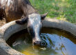
The submitted tumours originated from a wide range of anatomical locations, with tumours of the skin and subcutaneous tissue being the most frequently submitted. These were followed by tumours of the mammary gland tissue, uterus, and lymphatic tissue. Thyroid tumours were also represented. Neoplastic changes in other organs were submitted much less frequently (Fig. 1). Since tumours of the skin and subcutaneous tissue are easily noticed by owners during handling and grooming of the animals, this is presumably one of the main reasons why neoplasms at these sites were most frequently submitted. In contrast, pathological processes affecting internal organs are not necessarily detected by the owner or may require more complex surgical interventions.
Tumour Diagnoses
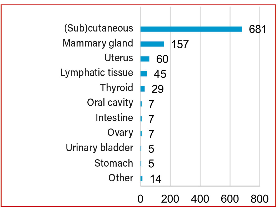
Lipoma was the most frequently diagnosed tumour in guinea pigs, followed by hair follicle tumours. Numerous other tumour types were also identified (Fig. 2). Given that the majority of samples originated from the skin and subcutaneous tissue, it is not surprising that tumours of adipose tissue and hair follicles were the most commonly diagnosed.
Frequency of Benign and Malignant Tumours
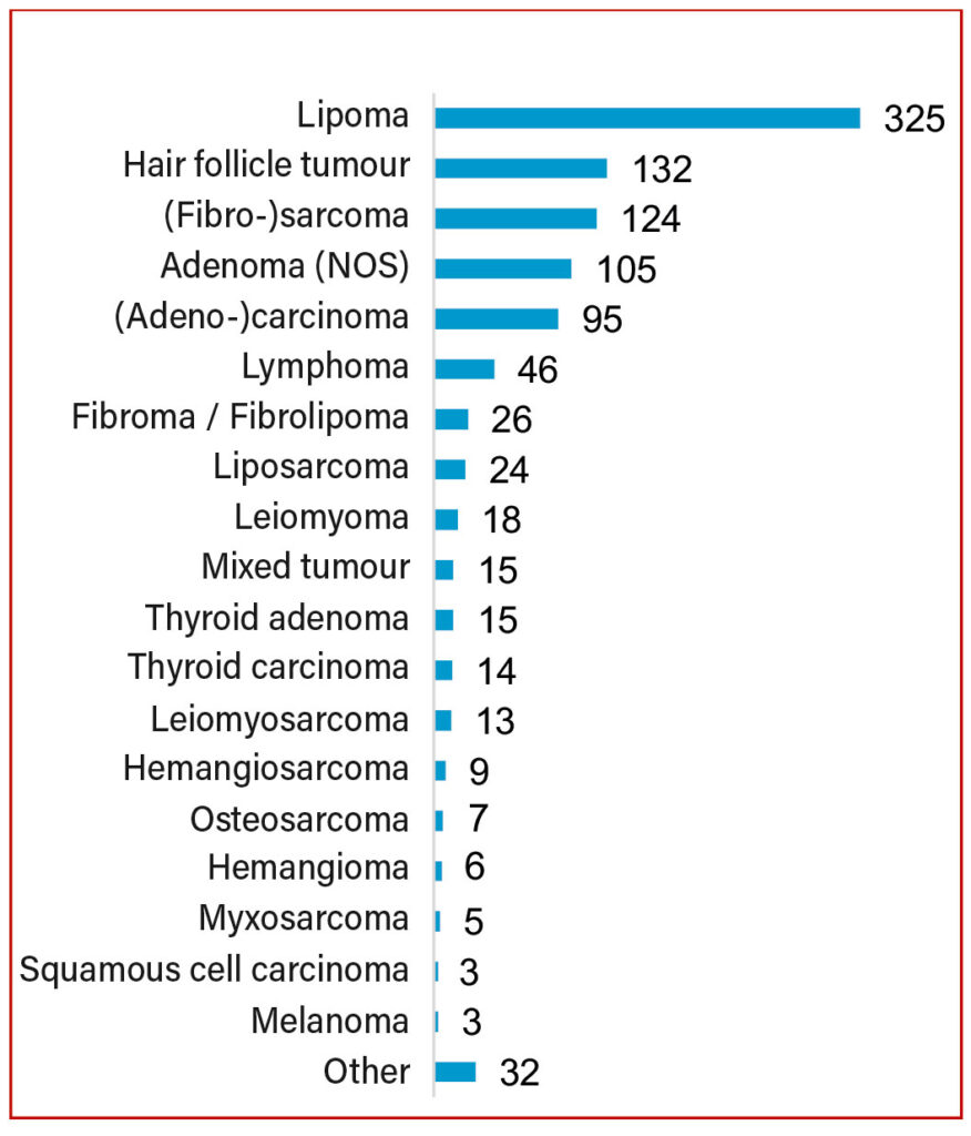
To provide a clearer overview of the ratio of benign to malignant tumours, this is illustrated schematically for the most frequently submitted tumour types (Fig. 3).
The most frequently occurring mesenchymal and epithelial tumours of the skin and subcutaneous tissue were further analysed to provide a clearer overview and to assess whether benign or malignant neoplasms predominated.
Mesenchymal Tumours of the Skin and Subcutaneous Tissue
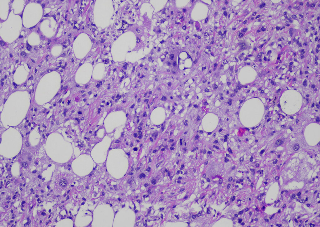
In guinea pigs, mesenchymal tumours predominated in the skin and subcutaneous tissue. A total of 508 mesenchymal tumours were diagnosed, of which 365 were benign and 143 were classified as malignant. The tumours included 325 lipomas, 13 fibromas, 13 fibrolipomas, and 6 haemangiomas. An additional 8 benign tumours were identified. Furthermore, 80 sarcomas were submitted, although a clear determination of the cell of origin was not possible. In addition, 29 fibrosarcomas, 24 liposarcomas (Fig. 4), and 10 other malignant mesenchymal neoplasms were diagnosed.
Epithelial Tumours of the Skin and Subcutaneous Tissue
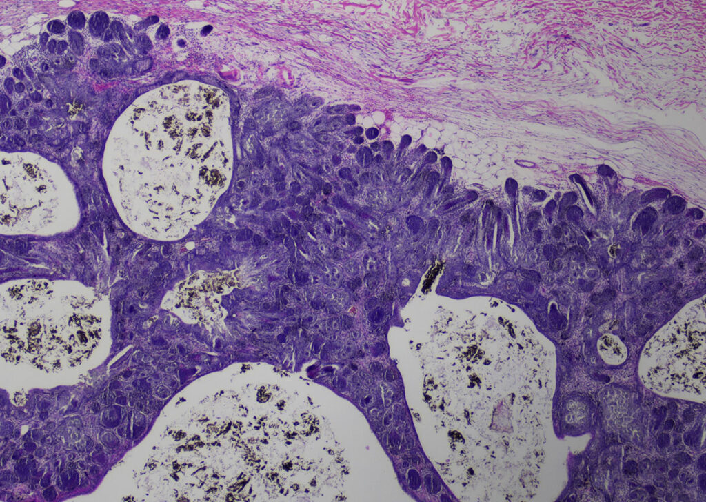
A total of 157 epithelial tumours were submitted, of which 142 were benign and 15 were malignant. The benign tumours comprised 124 trichofolliculomas (Fig. 5), 5 trichoepitheliomas, 3 pilomatrixomas, 3 adenomas, and 7 other benign neoplasms. The malignant tumours included 5 adenocarcinomas, 5 unclassified carcinomas, 3 squamous cell carcinomas, and 2 sebaceous gland carcinomas.
The other tumours of the skin and subcutaneous tissue included 11 cutaneous lymphomas, 3 melanomas, 1 fibropapilloma, and 1 carcinosarcoma.
-

-
Fig. 1: Tumour localisation in guinea pigs from Laboklin submissions (2013–2020)
Image source: Laboklin
-
-
Fig. 2: Tumour diagnoses in guinea pigs from Laboklin submissions (2013–2020). *NOS: not otherwise specified
Image source: Laboklin

-

-
Fig. 3: Distribution of benign and malignant tumours across the most frequently submitted anatomical sites
Image source: Laboklin
-
-
Fig. 4: Liposarcoma (H&E staining, 100× magnification)
Image source: Laboklin

-
-
Fig. 5: Trichofolliculoma (H&E staining, 20× magnification)
Image source: Laboklin

-
-
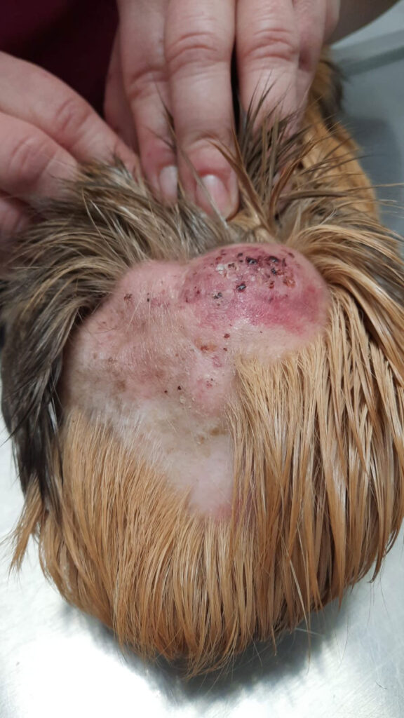
Fig. 6: Clinical presentation in a female guinea pig: ulcerative-crusted nodular proliferation of a cutaneous epitheliotropic lymphoma
Image source: © Kleintierpraxis Dr G. Raila

Lymphomas
Lymphomas also occur regularly in guinea pigs. In addition to lymphoma, a form of leukaemia has been described, particularly affecting young adult animals (under 3 years of age). These animals usually die within a few weeks.
The 46 lymphomas included in this study were predominantly located in the lymph nodes (n = 31), but also in the skin (n = 11), internal organs (n = 3), and the eye (n = 1).
A special form of cutaneous lymphoma is the cutaneous epitheliotropic lymphoma (syn. mycosis fungoides). This is a progressive tumour disease, characterised by infiltration of tumour T lymphocytes (memory T cells) into the epidermis and adnexa (Moore & Olivry, 1994). The aetiology of this disease is not yet fully understood. This form of lymphoma also occurs in guinea pigs and is often not initially recognised as a tumour. Clinically, erythema, alopecia, and scaling may be observed, often leading to an initial suspicion of a skin disease. Nodular proliferations may only develop at later stages of the disease (Fig. 6).
Mammary Tumours
In contrast to other animal species, mammary tumours are also regularly observed in male guinea pigs (Table 1) (Schöniger et al., 2025). During the study period, 157 tumours were submitted, of which 75 originated from female animals and 58 from male animals; the sex of 24 animals was unknown. A total of 78 adenomas, 7 fibroadenomas, 71 adenocarcinomas, and 1 undifferentiated carcinoma were diagnosed.
Table 1: Distribution of mammary tumours by sex
| Diagnosis | Number | F | FS | M | MS | U |
| Total | 157 | 74 | 1 | 44 | 14 | 24 |
| Benign | 85 | 47 | – | 17 | 7 | 14 |
| Malignant | 72 | 27 | 1 | 27 | 7 | 10 |
Legend: F: female, FS: female spayed, M: male, MS: male castrated, U: sex unknown.
Uterine Tumours
In contrast to rabbits, guinea pigs exhibit a wide range of uterine changes, which can be non-neoplastic or proliferative. Simple benign or malignant tumours may occur, and mixed tumours can also be diagnosed (Laik-Schandelmaier et al., 2017).
Between 2013 and 2020, 60 tumours from the uterine region were submitted, of which 37 were benign and 23 malignant. The tumours were classified as 28 epithelial, 27 mesenchymal, and 5 mixed tumours. Among the epithelial neoplasms, 19 adenomas were observed compared with 9 adenocarcinomas. Among the mesenchymal tumours, 17 leiomyomas, 8 leiomyosarcomas, and 2 undifferentiated sarcomas were identified. Of the mixed tumours, only one was benign, whereas four were malignant.
Thyroid Tumours
In contrast to other small mammals, guinea pigs frequently develop thyroid tumours. During the study period, 15 adenomas and 14 carcinomas were diagnosed.
Conclusion
In the present study of spontaneous tumours in guinea pigs, benign neoplasms of the skin and subcutaneous tissue predominated. Lipomas were by far the most common tumours, followed by trichofolliculomas. Cutaneous epitheliotropic lymphomas can be clinically difficult to distinguish from inflammatory skin conditions.
Mammary tumours occurred in both female and male guinea pigs. In this study, castrated females and males were less frequently affected than intact animals.
Histopathology is an important tool for the prognostic assessment of tumours, as neoplasms of the mammary gland, uterus, and thyroid can be either benign or malignant.
This study demonstrates that a tumour diagnosis in guinea pigs does not necessarily constitute a death sentence. In many cases, the underlying lesion is benign, and timely veterinary intervention with removal of the mass allows the animal to continue a normal, healthy life.
Dr Claudia Schandelmaier
Services offered
– Histopathology
– Cytology