A leukocytosis is defined as an increase in the number of white blood cells (leukocytes) in the blood.
Leukocytoses are relatively unspecific and detection should always be supplemented with a differential haemogram to classify the leukocyte population.
Leukocytoses can be divided according to the following causes:
- Physiological leukocytosis
- Stress leukogram
- Inflammation/infection
- Leukaemia
Essential components of a correct diagnosis and individual work up include:
- Detailed history and clinical exam including any previous testing
- Complete blood count (CBC) including a differential blood count
- Where applicable a morphological examination by a specialist (haematologist)of a freshly prepared blood smear
- Follow-up differential blood count (each blood count is only a snapshot)
- Interpretation in correlation with the clinical signs (“never treat a laboratory result, treat the patient”)
Physiological leukocytosis
The catecholamines adrenalin and noradrenalin trigger a physiological leukocytosis during the fight or flight reaction. Changes in the differential blood count caused by catecholamines are mostly seen in cats, horses, and juvenile animals. They typically involve minimal neutrophilia (mature neutrophils with no left shift) and lymphocytosis. The neutrophilia is caused by a shift of neutrophils from the marginal pool into the circulating pool. The lymphocytosis is caused by release of lymphocytes from the spleen into the peripheral blood. These changes are transitory and generally disappear within 30 minutes (after the animal has calmed down).
Stress leukogram
A stress leukogram is caused by endogenous (acute/chronic, often sick animals, hyperadrenocorticism) or exogenous (treatment) corticosteroids. Common changes are neutrophilia, monocytosis, lymphopenia, and eosinopenia. No every animal will have all of these changes at the same time. The most commonly observed changes are neutrophilia and lymphopenia. Monocytosis is mostly seen in dogs, in individual cases in cats and only rarely in horses and cattle.
As is the case in physiological leukocytoses, neutrophilia is caused by a shift of neutrophils from the marginal to the circulating pool. Additional neutrophils are also released from the bone marrow. Lymphopenia is caused by a variety of mechanisms, e.g. reduced release from lymph nodes as well as increased apoptosis as a result of high steroid concentrations. A stress leukogram is usually also a transient change, but can persist in cases of chronic stress or disease.
Inflammation/Infection
Inflammation is the most common cause of changes in the differential blood count. This is especially true of systemic inflammation and is only rarely seen in local inflammation. The severity of the leukocytosis and the differential blood count vary depending on the source of the inflammation or the infection. Typical changes in the differential are neutrophilia with a left shift. The mature and juvenile (band) neutrophils are released from the bone marrow into the peripheral blood. In general, the higher the proportion of band neutrophils, the stronger the inflammatory stimulus has been. Morphologically, toxic changes (Döhle bodies, foamy/vacuolated/basophilic cytoplasm) may occur.
An increased number of monocytes are often seen in chronic disease or when a disease is abating. Thrombocytosis also often has an inflammatory cause.
Causes of inflammation can be infections with bacteria, viruses, protozoa, or fungi, as well as immune mediated processes, neoplasms, necrosis, or foreign bodies.
Leukaemia
Leukaemia is a malignant disease of the bone marrow characterized by a neoplastic proliferation of myeloid or lymphocytic blood cells and, depending on the form, displacement of physiological haematopoiesis. Treatment is largely dependent on the form and type of leukaemia.
In contrast, a lymphoma is a clonal proliferation of lymphocytic cells that occurs in extramedullary tissues (mostly lymph nodes, lymphatic tissues, outside of the bone marrow). Lymphomas can be divided into stages 1-5.
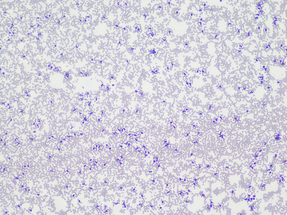
- Fig. 1: Canine blood smear, leukocytosis, 100x magnification, Diff-Quick stain.
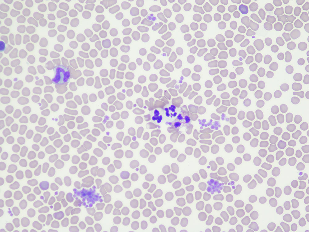
- Fig. 2: Canine blood smear with two band neutrophils and an eosinophil, small thrombocyte aggregates, 500x magnification, Diff-Quick stain.
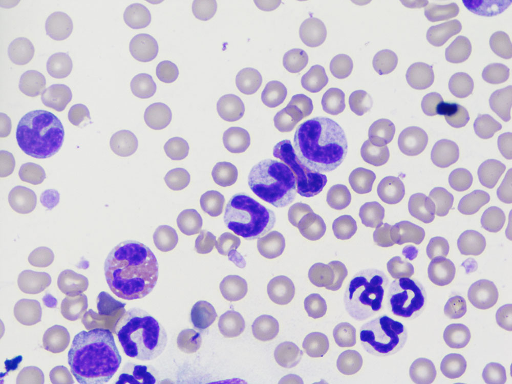
- Fig. 3: Canine blood smear with two band neutrophils and one metamyelocyte, with a segmented eosinophil in between, 1000x magnification, Diff-Quick stain.
- Fig. 4: Feline blood smear with chronic lymphocytic leukaemia arising from cytotoxic lymphocytes (T-cells), 500x magnification, Diff-Quick stain.
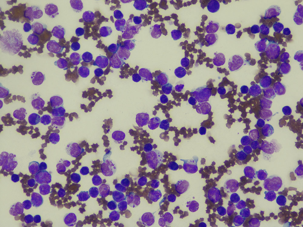
- Fig. 5: Canine blood smear, undifferentiated leukaemia, 500x magnification, Diff-Quick stain.
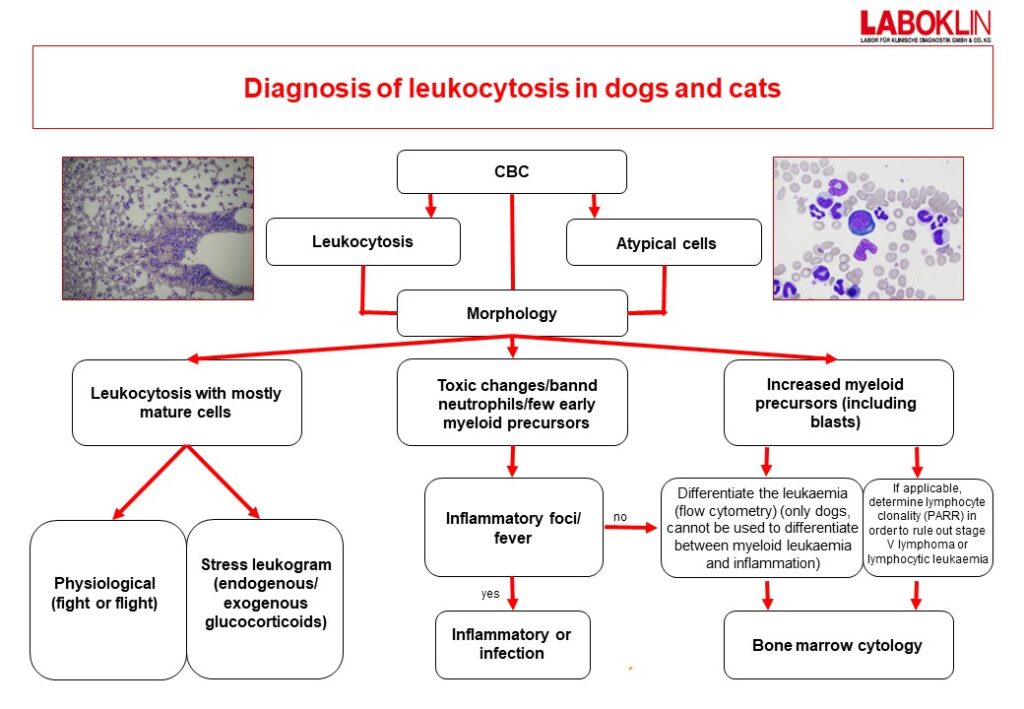
- Fig. 6: Diagnosis of leukocytosis in dogs and cats
Neoplastic cell proliferation in the blood can be categorized as follows:
- According to the clinical course of disease (acute, chronic, lymphoma with leukemic phase)
- According to the type of cell affected (lymphocytic, myeloid)
- According to the numbers of malignant cells in the peripheral blood (aleukemic, subleukemic, leukemic)
The most common acute leukaemia in dogs is usually myeloid. Myeloid leukaemias – often caused by the feline leukaemia virus (FeLV) – are also the most common form of acute leukaemias in cats. Horses are equally likely to be affected by lymphocytic and myeloid acute leukaemias.
In contract to acute leukaemias, chronic leukaemias are generally lymphocytic. Chronic myeloid leukaemias are rare in animals.
Cases in which leukaemia is suspected should be worked up in the practice and in the laboratory in the following steps:
- Detailed history including previous test results
- CBC including differential blood count
- Morphological examination by an expert (haematologist) of a blood smear prepared fresh in the practice
- If applicable, rule out ehrlichiosis, leishmaniosis in dogs and FeLV and FIV in cats
- If applicable, cytological examination of bone marrow or lymph nodes (depending on the case)
- In dogs with suspected stage V leukaemia or lymphoma: PARR and flow cytometry on EDTA blood
- In cats with suspected stage V leukaemia or lymphoma: PARR on EDTA blood
- In dogs and cats with suspected I-IV lymphoma: PARR on lymph node aspirate or lymphatic tissue.
