Particularities of the equine digestive tract
The horse’s digestive system is designed for the best possible utilisation of plant food. Digestion already begins with grinding the feed in the mouth and mixing it with saliva. Healthy teeth are a prerequisite for this. Horses are susceptible to oesophageal obstruction (choke) if they have dental problems, eat too fast or eat unsuitable food. Horses eat permanently, but their stomach is rather small compared to the size of the animal and only has a capacity of 8 – 15 litres, which means that only small amounts can be eaten at any given time. The pH of 2 is very acidic. Gastric acid is constantly produced. The pH value rises and hyperacidity is prevented when food is taken in and mixed with the mash. This reduces the risk of gastric ulcers. Enzymatic digestion and nutrient absorption takes place in the small intestine. This is where the breakdown of carbohydrates, proteins and fats occurs.
The large intestine consists of the caecum, colon and rectum. In the caecum, fibres that are difficult to digest, such as cellulose and pectin, are metabolised by numerous microorganisms into short-chain fatty acids, which serve as a source of energy to the animal. Microbial colonisation is determined by the type of feed. If it gets out of balance, it will result in faulty fermentation. In the colon, the formation of water-soluble B vitamins and vitamin C takes place as well as the absorption of liquid and electrolytes.
Clinical signs of digestive disorders
There are many general signs of gastrointestinal tract disorders in horses.
These include, among others:
• diarrhoea
• constipation, very dry faeces
• colic: abdominal pain, stomping, kicking stomach, tail swishing, frequent rolling, sweating, restlessness, apathy
• loss of appetite
• defaecation problems
• excessive gas
• poor performance, bad rideability
• flehmen, increased yawning
• choke, especially in older horses
There are many different causes of diarrhoea. Intestinal bacterial infections can, for example, induce hypersecretion of fluid into the intestine, which leads to the discharge of watery fluid before or after defaecation in the form of faecal liquid. Malabsorption can also promote diarrhoea. If less electrolytes and fluids are absorbed by the intestinal mucosa due to viral, bacterial or parasitic infections, osmotic imbalance may be caused. This increases the fluid content of the intestinal digesta and softens the faeces. Gastrointestinal symptoms can also manifest themselves as changes in intestinal motility. The equine digestive system bends and becomes narrow in many places. Hard, dry faeces can significantly increase the risk of obstruction. Faulty fermentation causes excessive gas which can lead to the displacement of intestinal sections.
The faecal sample – diagnostic possibilities
Bacteria:
Toxigenic strains of Clostridium perfringens and Clostridium difficile, salmonella, Lawsonia intracellularis and Rhodococcus equi are considered to be primarily pathogenic.
Although clostridia can be grown in anaerobic culture, the clinical signs are usually caused by the toxins. Therefore, toxin detection by means of enzyme-linked immunosorbent assay (EIA) is diagnostically more valuable than time-consuming cultivation, especially since clostridia are also part of the healthy intestinal flora.
Salmonella causes severe, feverish diarrhoea in horses. Foals also suffer from systemic diseases. In adult animals, asymptomatic shedding of the pathogen is possible. Sources of infection are mainly feed and water contaminated by faeces of infected birds, farm animals and rodents. Salmonella can be grown on special culture media or detected by PCR. It should be noted that they are not continuously shed. At Laboklin, testing for salmonella is always part of the bacteriological faecal analysis.
Lawsonia intracellularis, a gram-negative, obligate intracellular bacterium, is the causative agent of equine proliferative enteropathy (EPE). Suckling foals and especially weanlings are affected. Sick animals may suffer from a poor general condition, diarrhoea and colic symptoms, but often “wasting” is the only sign. Detection is done by PCR from faeces.
Rhodococcus equi causes severe pneumonia in foals. Additionally, intestinal disorders can occur. The pathogen can be detected by culture and PCR from tracheobronchial secretion (TBS) or faeces. PCR is more sensitive. Because of interfering factors which may be present in faeces, TBS should be preferred as sample material in this case.
Autovaccines:
Before the production of an autovaccine, a bacteriological examination must be carried out to isolate gram-negative pathogens. An oral vaccine is prepared from the inactivated bacteria and is administered orally to the horse for 20 days. This will help stimulate the production of secretory IgA in the mucous membrane. The use of autovaccines has been particularly successful in treating chronic digestive disorders, especially faecal water.
Viruses:
Rotavirus: Particularly in foals, it plays a role as a diarrhoeal pathogen. Typically, several foals contract the disease within a short period of time. Diagnosis is made by EIA from faeces.
Coronavirus: Mainly adult animals fall ill. Often, fever is the only sign. Morbidity is high but the mortality rate is low. Detection is carried out by PCR from faeces.
Parasites:
Parasitological faecal examinations should not only be carried out in case of digestive disorders, but also at regular intervals in horses that show no clinical signs. To increase the sensitivity of detection, it is recommended to test 3-day pooled faecal samples.
There are 2 different test methods available for the diagnosis of strongyles: Flotation provides a semi-quantitative result. Depending on the number of eggs per field of view, the quantity is indicated as low, moderate and high. In the modified McMaster technique, a defined amount of faeces is floated in a counting chamber so that the parasite stages present can be counted under a microscope. Here, the result is the number of eggs per gram of faeces. This is necessary if the horses are selectively dewormed.
Eggs of large and small strongyles cannot be distinguished microscopically. If they should be differentiated, a larval culture must be established.
Parascaris spp. is the most important endoparasite in foals and yearlings. In adult animals, patent infestation is rare. Detection is performed by microscopy after flotation.
Coproscopic detection of tapeworms only has a low sensitivity. The use of combined sedimentation-flotation techniques can increase the detection rate, but the intermittent shedding of eggs remains problematic.
Serum EIA is superior to faecal pathogen detection due to its higher sensitivity. This can be particularly useful for diagnosis at stock level.
Strongyloides westeri mainly occurs in foals up to 6 months of age; occasionally, adult horses are also affected. The eggs can be detected in fresh faecal samples by means of flotation. If the faecal sample is already several hours old, detection is carried out using the Baermann-Wetzel method.
Protozoa usually only lead to diseases in foals:
Cryptosporidia can be diagnosed by EIA or in a faecal smear stained with carbol fuchsin. Eimeria leuckarti and giardia can be detected microscopically after enrichment – however, for the detection of giardia, EIA and PCR have a higher sensitivity.
Sand:
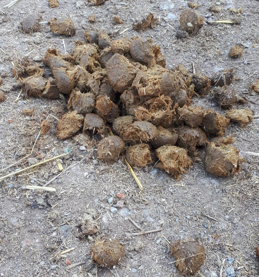
When hay is fed on sand paddocks or if there is insufficient pasture growth, there is a chance that horses take up too much sand when eating – the risk of sand colic increases. Faecal sand can easily be detected directly at the stable or in the practice. To do this, the horse droppings are dissolved in water, e.g. in an examination glove. Sand settles and can be seen on the bottom. A positive detection of sand is conclusive. However, if no sand is excreted, the presence of sand in the digestive tract cannot be excluded.
Outlook:
Drinking water for animals:
Aside from infectious diarrhoeal pathogens, there are many other causes that can lead to digestive disorders. Monitoring the food and water quality should not be forgotten. Especially if animals do not have access to drinking water and instead drink from open water points or wells, water quality should be tested at regular intervals. At Laboklin, a drinking water profile particularly tailored to the needs of horses will soon be available.
Microbiome:
As in all other mammals, the equine intestinal microbiome also has a close functional connection to the host. A well-functioning digestive system with a well-established intestinal bacterial flora is therefore of essential importance for equine health. Although the causal relationships are still subject to current research, intestinal microbial homeostasis is strongly influenced by factors such as digestive disorders or feed changes. Initial studies on the characterisation of dysbiotic changes of the intestinal microbiome in horses already show promising results. For example, certain members of proteobacteria seem to be significantly overrepresented in horses with faecal water.
In contrast, anaerobic clostridia species and members of the phylum Verrucomicrobia are considerably reduced. In the future, targeted testing of the intestinal microbiome could help to diagnose shifts in the intestinal microbiota and identify predispositions for gastrointestinal disorders. Based on this, it would be possible to prevent or treat gastrointestinal disorders already before the onset of clinical signs by using coordinated treatment concepts (e.g. selected pre- and probiotic agents).
Ann-Kathrin Schieder, Dr. Ronnie Gueta


