Overview and data situation
Apart from orthopaedic problems, intestinal diseases are the most frequent causes for consultations in the veterinary practice. Although primary tumours are rare, they do occur. The most encountered tumours are lymphomas, spindle cell tumours and adenocarcinomas.
For clinicians, it can be a diagnostic challenge to distinguish between chronic enteritis, space occupying inflammatory processes and intestinal or extraintestinal neoplasias, as the clinical symptoms are non-specific. In addition to weight loss and acute colic, recurrent colic and, less frequently, anorexia, diarrhoea or fever also occur.
Intestinal tumours can occur in all breeds, there seems to be no disposition. In one American study on intestinal tumours in horses, a breed predisposition for Arabians was described, but since this breed accounted for almost half of the animals in the study, the clear dominance of Arabians may reflect overrepresentation of this breed in the American horse population.
Unfortunately, it is not possible to make a macroscopic statement regarding the type of circumferential increase or its dignity. Tumours, both benign and malignant, can vary greatly in size. A histological examination is therefore essential to determine the origin and tumor behaviour. It is important to always send in the whole tumour after removal, if possible, in order to make a statement that is as reliable as possible.
The patient’s lymph nodes should also be resected, if necessary, because this is a decisive factor in assessing the prognosis.
Tumour types, their characteristics and prognoses
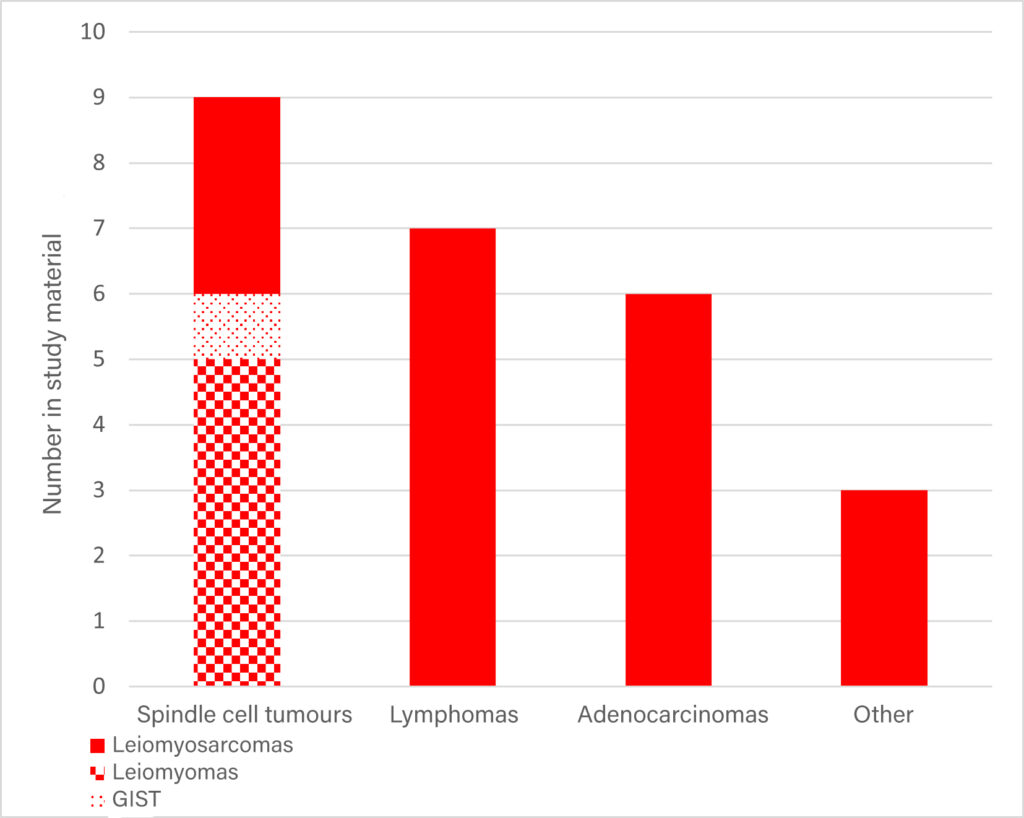
The “classic” intestinal tumours in horses are the spindle cell tumours, the lymphomas and the adenocarcinomas, with the lymphomas making up the majority in the literature. There are few major case collections. In an internal study at Laboklin, 34 cases from 2011 – 2023 were collected from routine diagnostics that fulfilled the criterion of direct association with the intestine. 25 of these were classified as tumours (Figure 1), the remaining were reactive or inflammatory processes.
Spindle cell tumours
Spindle cell tumours are tumours of mesenchymal origin. The most frequent location is the small intestine. To differentiate between benign and malignant mesenchymal tumours, infiltrative growth is the most important diagnostic criterion. It can be further differentiated into various types, some of which can only be distinguished from each other by means of immunohistology.
In our study material, spindle cell tumours made up the largest proportion, where the ratio of benign leiomyomas to malignant variants (leiomyosarcomas, gastrointestinal stromal tumours (GIST)) was approximately 1:1. All leiomyomas and two leiomyosarcomas were found in the small intestine, one leiomyosarcoma in the colon and the GIST in the caecum.
In horses with leiomyosarcoma in the small intestine, locally invasive growth does occur, but no metastasis has been described in the literature at this time. It is therefore debatable whether a complete removal of the tumour with sufficient distance to the healthy tissue in the region of the small intestine can be considered curative. The situation is apparently different with leiomyosarcomas of the caecum, where metastases in the liver or peritoneum could be detected and these are therefore to be assessed as having a poorer prognosis.
Lymphomas
Lymphoma is the most common intestinal tumor in the horse and the intestinal form is, after the multicentric and the cutaneous form, the third most common localisation with 11 %.
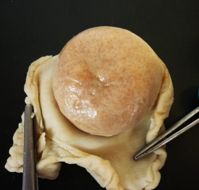
The most frequent location for lymphomas is also the small intestine (Figure 2), which has been confirmed both in the literature and in our study material. A multicentric occurrence involving lymph nodes or the spleen and other organs is also possible and can be proven if the material submitted is appropriate.
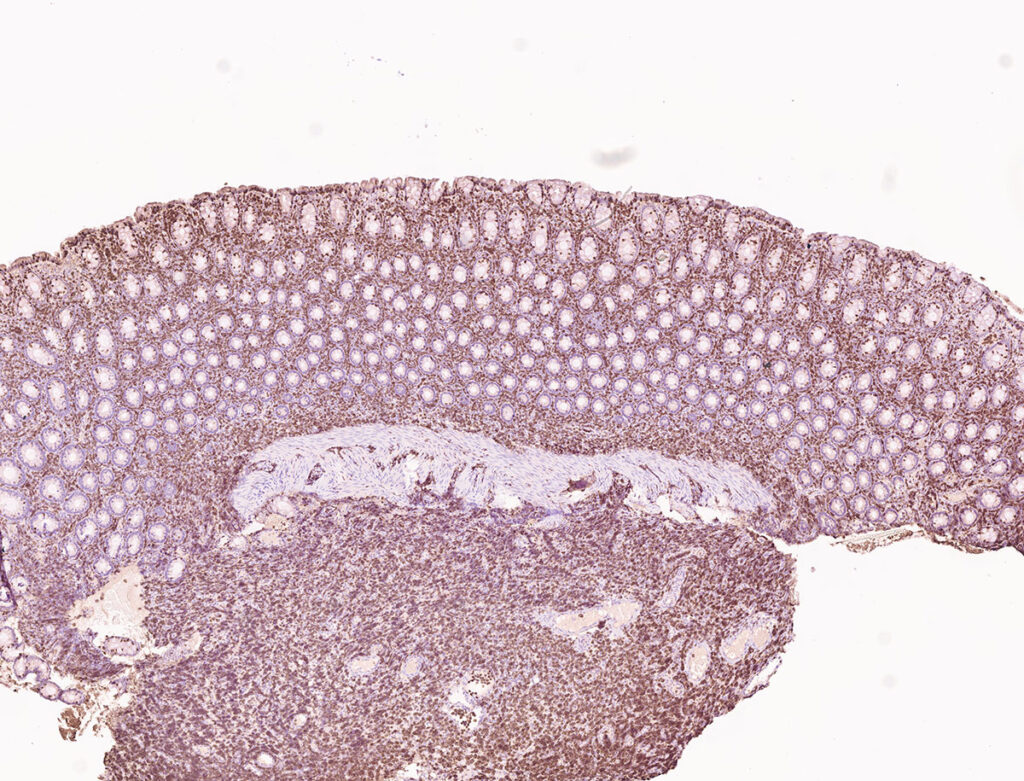
In some cases, especially in the case of transmural growth, the diagnosis of lymphoma can already be made on small deep biopsies (up to 0.5 cm). The growth behaviour in depth is a helpful indication for the evaluation of malignancy and differentiation between a reactive and a neoplastic process. Since the deeper parts of the intestine are often not present in intestinal biopsies, the lymphocyte population must be identified solely on the basis of their morphology, the mitotic count, which is often very low, and the results must be evaluated in the light of the preliminary clinical report. Thus, this less invasive method is a challenge for pathologists, as the predominant type of lymphoma in horses is small-cell, mitotic and well-differentiated and sometimes difficult to distinguish from reactive lymphocyte infiltrates on the basis of biopsies. It should be noted that in horses there is usually no leukaemia and other lymphatic organs (e.g. peripheral lymph nodes) are often not enlarged. Furthermore, clonality testing which is often used to distinguish rective from neoplasic proliferations of lymphocytes in small animals is not available for horses, immunohistological examination is often applied in such cases, which can contribute decisively to the diagnosis in many cases through B- and T-cell differentiation. Both in the in-house study and in the literature, most cases are T-cell lymphomas (Figure 3). One B-cell lymphoma in the study material showed an amyloid deposition as a special feature, which was also detectable in the tributary lymph node.
Basically, intestinal lymphomas in horses have an unfavourable prognosis. The median survival time depends on histological/ immunohistological type and is given in a recent study with a median of 60 days (Bacci et. al., 2020).
However, there was also one horse that had a survival time of 650 days at the time of publication.
- Image source: envatoelements
-
Fig. 1: Distribution of tumour types in the own examination well 2011 – 2023
Image source: Laboklin
-
Fig. 2: T-cell lymphoma in the small intestine of a 2-year-old warmblood gelding
Image source: Laboklin
-
Fig. 3: Immunohistochemical findings of a T-cell lymphoma from biopsy material
Image scource: Laboklin
-
Fig. 4: Macroscopy of a mandarin-sized adenocarcinoma in the colon of a 15-year-old pony gelding
Image source: Laboklin
-
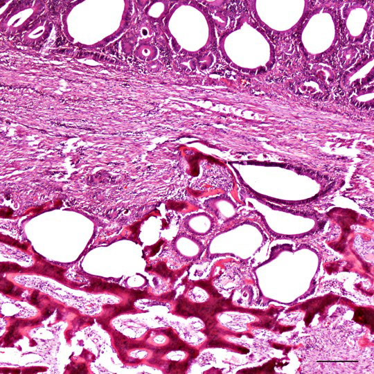
Fig. 5: Histology of an adenocarcinoma with osseous metaplasia
Image source: Laboklin
Adenocarcinomas
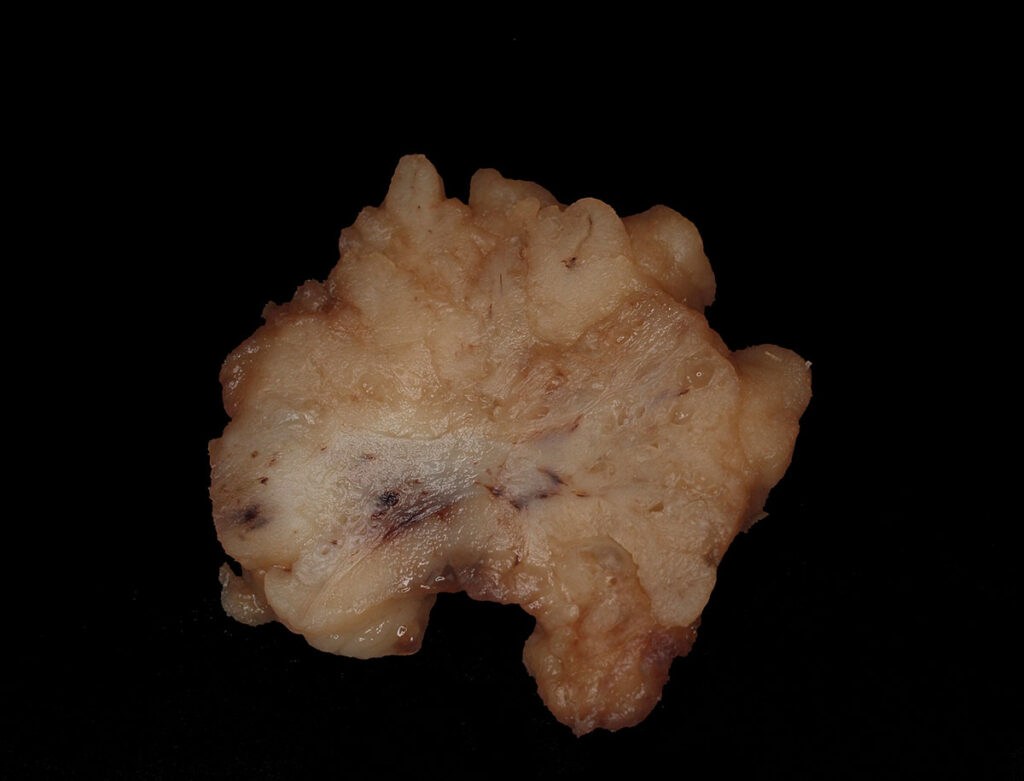
There is contradictory information in the literature on the localisation of adenocarcinomas, with reports of dominance in both the small and large intestine (Figure 4). In our study material, no clear trend with regard to distribution could be detected. Histologically, they are characterised by an infiltrative growth and more frequently show osseous metaplasia (Figure 5).
Intestinal adenocarcinomas are generally considered to have an unfavourable prognosis and tend to metastasise in addition to infiltrative growth. Nevertheless, there are reports according to which surgical removal has led to a survival time of three to five years after diagnosis up to complete healing without metastasis and recurrences and thus, similar to the spindle cell tumours, an early, complete resection can also lead to a longer survival time here (if there are no metastases or vascular infiltration to date).
Other tumours and circumferential growths
In addition to the “classic” tumours already listed, there are also rarer neoplasms in addition to non-neoplastic changes (haematoma, nodular inflammations). For example, nodular vascular proliferations occurred in our own examination material. These were angiomatoses in the colon and a lymphangioma in the small intestine.
Conclusion
Intestinal tumours in horses are rare, but must nevertheless be considered in the differential diagnosis of gastrointestinal problems.
It is not possible to draw conclusions about histogenesis and tumor behaviour from the non-specific clinical symptoms or macroscopy. Often the horses are only clinically noticed when the circumferential proliferations have reached a certain size and then, regardless of the tumor behaviour, they lead to colic with the obturations and passenger disturbances associated with the circumferential proliferation, as can also occur with other positional changes.
Neither size nor shape can allow conclusions to be drawn about histogenesis and tumor behaviour; further histological and, if necessary, also immunohistological examination is therefore inevitable.
Ultimately, until a final histopathological diagnosis is made for nodular changes, the only option is to resect the tumour as thoroughly as possible, including the lymph nodes. Depending on the results, however, this can already be curative or indicate an extension of the survival time.
Dr Lena Kempker

