Part I: BEVA Congress, September 2022, Liverpool
John Pringle (Swedish University of Agricultural Sciences): Strangles
The expert discussed some recent studies that address the detection of silent carrier horses. According to Pringle, a single examination of a guttural pouch lavage sample is not enough to rule out a possible carrier status. It is also an option to perform multiple nasopharyngeal washes if it is not possible to do several guttural pouch washes. After an outbreak, the prevalence of silent carriers varies between 3 and 35 %. Shortly after an infection, performing a guttural pouch lavage in horses with empyema is sufficient to identify carriers. If, however, carriers are identified many months after recovery, it seems that both a nasopharyngeal lavage and a guttural pouch lavage sample are required, as long-term carriers often have neither empyema nor chondroids. Thus, a purely visual examination is not sufficient to exclude a carrier.
It is important to note that there may be negative PCR results from guttural pouch lavage samples in these horses. Simpler methods, such as combining guttural pouch and nasopharyngeal lavage samples, could thus facilitate the detection of carriers in the future.
If a carrier status is suspected, testing for antibodies does not offer any advantages as many long-term carriers are seronegative or may not differ serologically from other members of the herd which have recovered after an outbreak. Furthermore, recent exposure does not correlate with antibody titres. Antibody testing is only recommended for the detection of seroconversion during an outbreak in order to assign horses to the correct quarantine groups (green, yellow, red). One way of preventing a severe course of the disease can be the marker vaccine which has recently become available on the market. Using a marker vaccine means that no false positive serological tests are to be expected and thus antibodies will only be detectable in a natural infection. The aim of the vaccination is to reduce clinical signs in acute infection as well as the number of abscesses. Moreover, it is possible to immunise healthy horses (aged 5 months and older) during an outbreak. A potential influence on the carrier status has not yet been proven, but the reduced number of abscesses could also prevent/reduce the potential shedding of pathogens. It is still debatable whether vaccinating a carrier is likely to be successful.
- Photo credits: envatoelements
-
Fig. 1: Meadow saffron
Photo credits: shutterstock
-
Fig. 2: Obese horse
Photo credits: envatoelements
Richard J. Piercy (RVC): Myopathies – PSSM 1/2 and atypical myopathy
1. PSSM 1/2
According to Piercy, if myopathy is suspected, it can initially be confirmed by detecting elevated serum or plasma activities of the muscle enzymes creatine kinase (CK) and aspartate aminotransferase (AST). However, (apparently) subclinical elevations of CK and AST are often found in horses with poor performance. It is usually difficult to assess the clinical relevance of these elevations. In some types of myopathies, CK and AST activities are actually within the reference range, even though the horses show clinical signs. CK and AST can be important indicator of the degree and timing of muscle damage. CK activity is highest 6 – 12 h after muscle damage and then decreases with a half-life of about 12 h. In contrast, AST activity is highest after about 24 h and may also remain elevated for several days to weeks. A CK activity of 10,000 IU/L indicates 400 – 500 mg of damaged muscle mass.
If exertional rhabdomyolysis is suspected, an exercise test should be performed. Unfortunately, there are no standardized protocols. Piercy recommends 20 minutes of light to moderate exercise (trotting) on the lunge and measuring CK and AST activities in advance, after 4 h and after 24 h. However, in horses with myopathy, the percentage increase in CK activity after exercise can vary greatly – in some horses, there may be no change at all even though there is severe muscle damage.
Polysaccharide storage myopathy (PSSM) is an exercise-induced congenital myopathy. Animals heterozygous for PSSM often have normal muscle enzyme activities. Homozygous animals, however, may have increased muscle parameters. A genetic test is available for the diagnosis of PSSM1. Equine malignant hyperthermia (EMH) and immune-mediated myositis can also be excluded by genetic testing. As far as PSSM2 or myofibrillar myopathy (MFM) are concerned, there is now information that contradicts the validity of the genetic tests available.
The diagnosis of PSSM2 can still only be confirmed by muscle biopsy and is based on amylase-resistant inclusions, myopathic muscle fibres and a negative genetic test for PSSM1. Desmin aggregates, on the other hand, confirm the diagnosis of MFM. According to Piercy, muscle biopsy is the best method for confirming equine motor neuron disease, sarcocystosis or myopathy in horses with occasional or mild increases in CK or AST activity and in horses showing other clinical signs (such as paresis) without elevated muscle enzymes. In exertional rhabdomyolysis, a muscle biopsy is not helpful as it only provides information about the severity and chronicity of the disease. However, in sport horses, a muscle biopsy may be of interest to assess the prognosis of rhabdomyolysis.
According to Piercy, non-specific myopathies with mild CK and AST elevations occur in Icelandic horses, Connemara ponies or warmblood horses.
His conclusion was: Atypical myopathies should be considered in horses with non-specific elevated CK and AST activities. For pathological examination, muscle biopsies should be sent unfixed and refrigerated in a plastic container to a specialised laboratory.
2. Atypical myopathy
The prevalence of atypical myopathy (AM) has increased over the past few years. The disease is caused by the toxin hypoglycin A (HGA) contained in the seeds and seedlings of the sycamore tree (Acer pseudoplatanus), which horses may ingest while grazing in autumn, spring and winter. The mortality rate is about 60 – 70 %. Horses with AM may show various signs of muscle pain, stiffness, weakness, head and neck hanging down, muscle fasciculations or tremors, breathing difficulties, lethargy, colic-like symptoms and, typically, myoglobinuria. Susceptibility to HGA varies between horses. This also means that horses that seem to show no clinical signs can have high levels of HGA in their blood and vice versa. According to Piercy, it was possible to detect HGA in the serum of horses with slightly increased CK activity (CK < 1000 IU/L) without any apparent cause. Subclinical cases are therefore possible, the only clinical sign may be poor performance. For the diagnosis of atypical myopathy, serum or plasma HGA as well as the metabolites can be determined. HGA is known to have a half-life of 2 days in blood. If large amounts of acylcarnitine are produced as metabolites, the survival rate of affected horses is significantly reduced.
S. Möller (Laboklin): Detection of colchicine in suspected meadow saffron poisoning
Meadow saffron poisoning is caused by the ingestion of tropolone alkaloids (e. g. colchicine) from leaves (spring), seed capsules (summer) or flowers (autumn) (Fig. 1). Horses often eat dried plant parts in hay. The intake of high doses of colchicine can lead to colic, bloody diarrhoea, circulatory disorders or even death. According to the literature, a dose of 0.17 mg/kg bw already leads to severe diarrhoea, while the lethal dose for horses is 1 mg/kg bw (per os).
The aim of the study was to develop a valid test procedure for the detection of colchicine poisoning. A total of 91 left-over urine samples from horses sent to Laboklin were analysed for cholchicine. 28 urine and blood samples (EDTA, serum) were analysed from a farm where horses became ill after ingesting contaminated hay. The horses suffered from recurrent colic of unknown cause, diarrhoea or faecal water, gastric ulcers, hypoproteinaemia, oedema as well as lameness of unknown origin. Colchicine was detected in all of the urine samples of the suspected horses (13.20 ± 32.12 ng/ml, max. 152.80 ng/ml). The blood samples, however, were tested negative for colchicine. This study shows that it is possible to detect colchicine in urine and thus support the diagnosis of meadow saffron poisoning.
Part II: AAEP Annual Convention, November 2022, San Antonio/Texas
During the “Kester News Hour”, a number of important publications from the past year were presented.
K. Thane et al.: Effect of varying sampling times on the results of the TRH stimulation test in the diagnosis of PPID
Conclusion: The second blood sample should be taken exactly 10 minutes after the TRH injection. Sampling after 9 or 11 minutes leads to approx. 10 % differences in the results and to incorrect interpretations in about 20 % of the cases.
N. Pusterla: Role of Sars-CoV-2 in the equine population
Horses are susceptible to the virus but do not develop clinical signs. They can, however, seroconvert. In a racing stable with a large number of sick jockeys, 3.5 % of the horses were seropositive, but none had a positive PCR result.
C. B. Fernandes: Behaviour and some perinatal parameters of mule foals
Worldwide, there are approx. 10 – 11 million mules, which are considered to be extremely hard-working, persevering, feed-efficient, intelligent and not easily scared. For mule foals, too, the first days of life are the most critical ones because of the change in foetal circulation and nutrition to pulmonary respiration and enteral feeding. Mule foals have a higher APGAR score than horse foals; they are faster at seeking the udder, standing and nursing. Mares with mule foals eliminate the placenta faster than mares with horse foals. However, meconium discharge is significantly later in mule foals: up to 72 hours p. p. Overall, mule foals have a faster neurological and hormonal adaptation to extra-uterine life (“hybrid vigor phenomenon”, “genetic improvement”). The gestation period until mule foals are born is the same as for horse foals.
L. Huggins: Retrospective evaluation of mares with abnormal behaviour and their endocrinological test results, especially the diagnosis of granulosa cell tumours
31,981 blood samples from mares with abnormal behaviour were included in the study. In 86 % of the mares, the endocrinological results were not suspicious. The sensitivities of the tests were 90 % for AMH, 80 % for inhibin B and 40 % for testosterone. Abnormal rectal examination findings as well as “stallion-like behaviour” in the clinical history, however, correlated with abnormal hormone concentrations.
Conclusion: If there are problems with mares showing undesirable behaviour, they are rarely found in the reproductive tract.
Equine Endocrinology Group: What is new?
Testing for PPID in horses without any signs is not recommended!
The reference ranges for ACTH were raised slightly. This leads to fewer positive but more borderline results. ACTH levels that are borderline do not allow a direct diagnosis of PPID, but these horses should be monitored further. If the ACTH levels of treated horses are still above the reference range, even though the clinical signs of the horses are much better, the dose does not automatically have to be increased! Instead, the patient’s clinical presentation should be checked more often. For borderline results where the clinical picture is also inconclusive, short-term diagnostic treatment should be considered.
For horses that do not respond well to pergolide tablets or do not tolerate them at all, a cabergoline preparation is available in the USA (human) which is injected once every two weeks and leads to an improvement in clinical signs. H. C. Schott reported on long-term treated PPID patients where he observed that adjustments in ACTH levels can still occur years after starting pergolide treatment. Overall, pergolide improves the quality of life, but not the life expectancy.
EMS: For the oral Karo Light corn syrup test, blood samples should be taken after 60 and/or 90 minutes. Insulin and glucose are determined. Alternatively, an insulin resistance test can be carried out. Fasting not required! How to proceed: baseline blood sample to determine glucose, immediately followed by injection of 0.10 IU/kg insulin.
2nd blood sample after 30 minutes: The glucose concentration should have decreased by 50 %.
It is also possible to first determine the insulin response to the usual food: Give the horses their normal food or let them graze for 5 – 6 hours. Collect a blood sample after 2 hours to determine the insulin level.
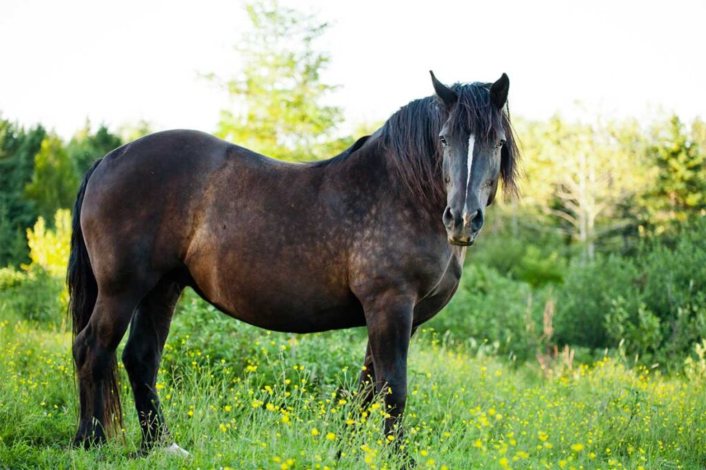
Obese horses (Fig. 2) with normal insulin: insulin regulation still works. Maybe determine leptin, but this is not yet commercially available.
T. Sundra et al. from Australia presented a very promising approach to the treatment of EMS. The group used ertugliflozin to treat hyperinsulinaemia and laminitis. It is a sodium-glucose co-transporter-2 inhibitor which promotes glucose excretion via the kidney. The study was carried out on 36 ponies. Treatment was initially given for 6 weeks, or longer in some horses. The results were promising: In all ponies, the insulin concentration was considerably reduced and the horses lost a lot of weight. Lameness also mostly disappeared with the radiographs of the horses being unchanged. No further episodes of laminitis occurred. Hepatic parameters and triglycerides should be monitored closely. Triglycerides were frequently elevated, but no horse developed hyperlipaemia. Some horses developed PU/PD during treatment. Long-term studies and controls are still lacking and the medicine seems to be very expensive. However, the results and images shown suggest that there is a promising approach to treating EMS.
Dr Antje Wöckener, Dr Svenja Möller


