Small pets are now among the most popular domestic animals in Germany (5 million small pets in 5% of all households, according to a survey by the IVH, the German Industrial Association of Pet Care Producers, in 2020). The willingness of owners to take their pets to a vet in case of illness has increased. As a consequence, more samples are sent to the laboratory for analysis.
In 2020, for example, 797 samples from small mammals were sent to Laboklin GmbH & Co. KG for histopathological examination.
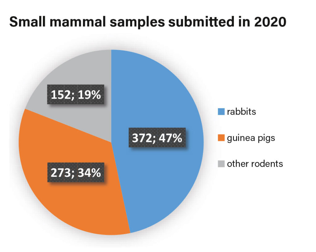
Most of the samples were from rabbits, closely followed by samples from guinea pigs (Fig. 1). Other species were grouped under the umbrella term “rodents”: rats (n=85; 10.7%), hamsters (n=26; 3.3%), gerbils (n=12; 1.5%), chinchillas (n=11; 1.4%), degus (n=8; 1.0%), mice (n=6; 0.6%), squirrels (n=3; 0.4%) and one alpine marmot (0.1%). Samples from ferrets (n=77) and hedgehogs (n=58) were not included in these statistics.
Below, the possibilities are discussed when and why a certain examination is useful and where there may be limitations.
Cytology
Cytology is a method used for the microscopic diagnosis of single cells. Fine needle aspirates (FNA) are suitable samples for examination. They may come from masses or even from organs. Other suitable samples are impression smears, e.g. from open wounds or scabs. Body cavity effusions can be analysed for their cell content, cell composition and the presence of pathogen structures.
Special stains are available as well. They are used, for example, to detect pathogen structures. Ziehl-Neelsen staining (ZN) detects acid-fast rods (e.g. mycobacteria). PAS reaction (periodic acid-Schiff reaction) is used, among others, to detect fungal elements. Prussian blue staining differentiates haemosiderin from other pigments through its blue colouration.
Advantages
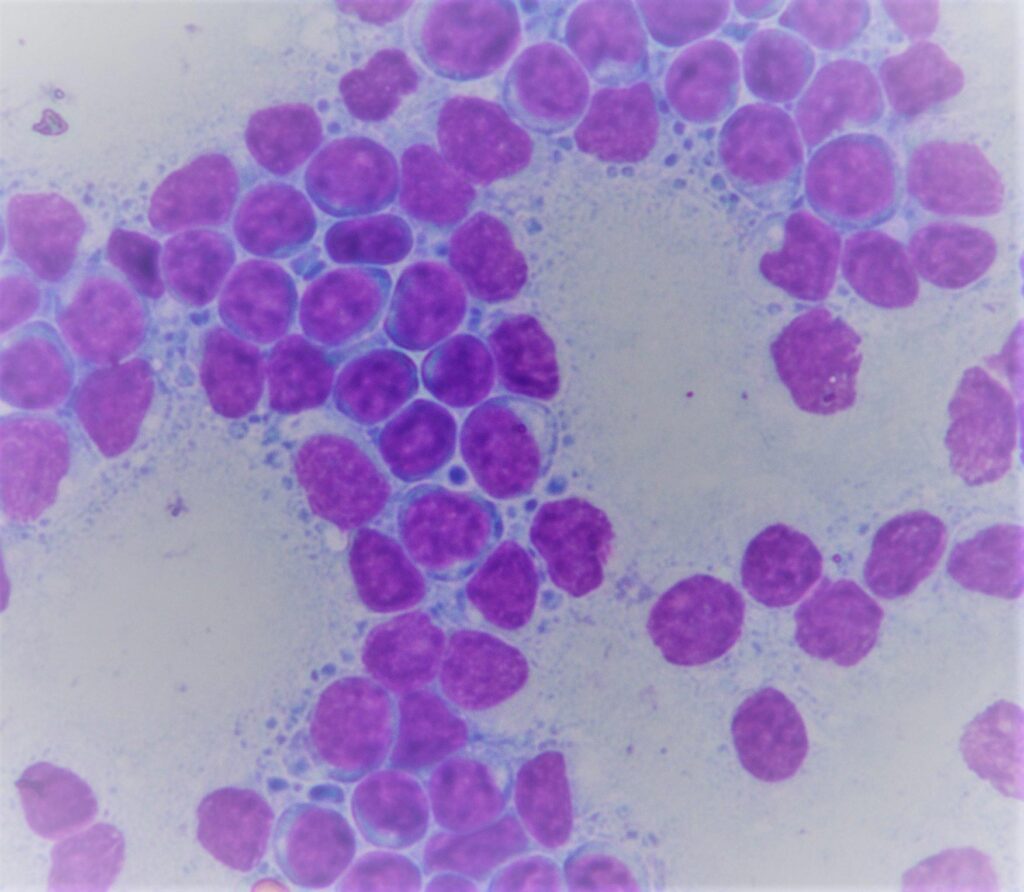
Cytological examination is a micro-invasive procedure. As small mammals are often more sensitive to anaesthesia and surgical intervention than other domestic animals, cytology is sometimes preferred to surgical extirpation. It is a simple type of test that provides quick results. With this method, it is often already possible to distinguish between an inflammatory and a tumorous process (Fig. 2).
Disadvantages/limitations
Only positive cytological findings are conclusive. This means a tumour can only be diagnosed if tumour cells are found in the preparation. If no tumour cells are present, it is not possible to definitely rule out a tumour. The clinical history is also important for cytological evaluation. Especially in cytology, which is based on the analysis of single cells, information on the sampling site, the clinical picture and previous treatment is often essential. In order to ensure diagnostically conclusive results, sufficient cell material must be obtained. This can sometimes be complicated, e.g. if there is a cystic mass or if the cells are arranged in a dense cell cluster making aspiration of cell material difficult. Diagnosis is hampered by slides covered with cover glasses or adhesive tape (insufficient staining of the cells, formation of air bubbles). Smears that are too thick also make diagnosis difficult. In these cases, it is no longer possible to evaluate single cells. Furthermore, cell quality suffers as it cannot be guaranteed that cells are sufficiently fixed by air drying.
Histopathology
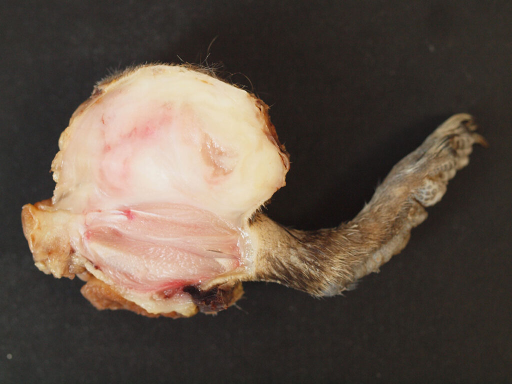
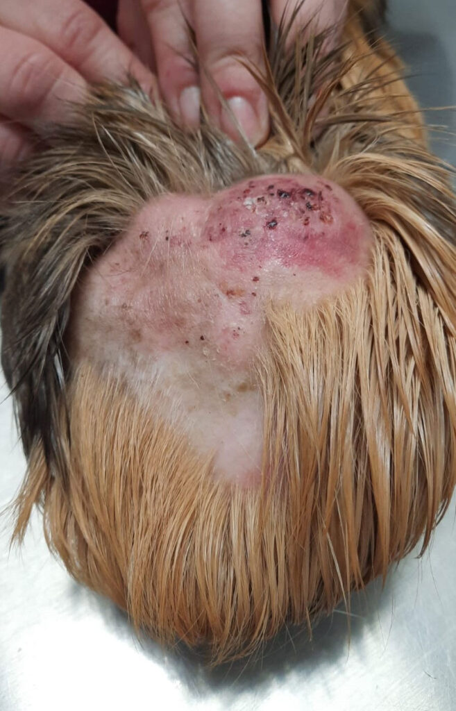
In contrast to cytology, histological examination involves the examination of a formalin-fixed tissue structure. It is thus possible to evaluate the organ structure (regular organ-specific or autologous growth in case of neoplasia). Masses, biopsies or organ samples from small mammals can also be sent in for examination (Fig. 3 and 4).
Advantages
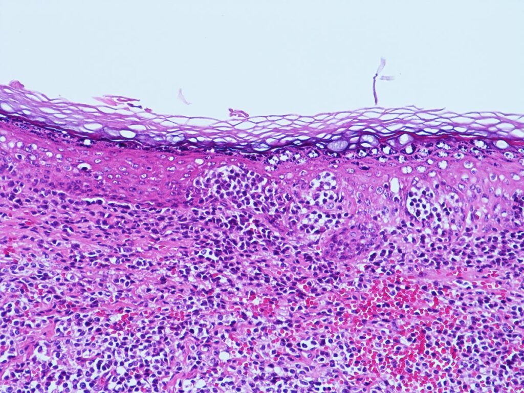
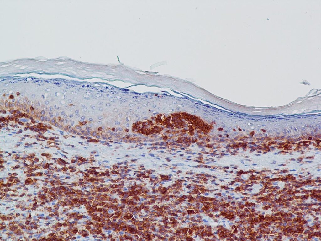
It is possible to evaluate whether the underlying process is inflammatory or tumorous. If there is a mass, this type of examination cannot only clarify whether it is a neoplasm but also checks for completeness, i.e. the resection margins are evaluated. The analysis of skin punch biopsies is also gaining importance in small mammals. Sometimes, histopathology is the only way to make a conclusive diagnosis, for instance in sebaceous adenitis in rabbits. It can only be determined histologically whether sebaceous glands are present or not. To clarify an underlying infectious cause, a variety of further special stains can also be used in histopathology, e.g. PAS reaction, ZN stain, Warthin-Starry (WS) stain (detection of spirochaetes, e.g. Treponema paraluiscuniculi) and many more. Immunohistochemistry, which has become a standard test for dogs and cats, is, in some areas, also established for small mammals. For example, at Laboklin, the lymphocyte markers CD3 (T lymphocytes) and CD79α (B lymphocytes) have been validated for guinea pigs, rabbits and ferrets (Fig. 5a and b). In all three species, lymphoma is diagnosed quite frequently. Immunohistochemistry can then be used for further differentiation into B- or T-cell lymphoma.
Disadvantages/limitations
As in cytology, it is important to provide the clinical history, i.e. information on the sampling site, the clinical picture and possible previous treatment, in order to evaluate the submitted sample. Moreover, immediate fixation of the sample is required (4% neutral buffered formaldehyde ≙ 10% formalin). If it is not done or if the quantity is not sufficient, autolysis of the tissue will occur which limits getting a reliable evaluation. The formation of artefacts, such as freezing artefacts or heating artefacts due to thermosurgery, may also be a disadvantage. Specimens should be submitted from representative sites. Samples for histology shall not be too small, as otherwise the tissue structure cannot be evaluated. Skin punch biopsies in particular should have a minimum diameter (approx. 0.4 cm), if site and species permit (if not, alternative sampling, e.g. “shave biopsy”). Limitations also arise if a secondary lesion, e.g. inflammation, overlaps the primary process (tumour).
-
Fig. 1: Pie chart of small mammal samples submitted in 2020
Photo credits: Laboklinn
-
Fig. 2: Guinea pig: FNA lymph nodes: lymphoma
Photo credits: Laboklin
-
Fig. 3: Degu: limb with mass: sarcoma
Photo credits: Laboklin
-
Fig. 4: Guinea pig: cutaneous epitheliotropic lymphoma
Photo credits: Kleintierpraxis (small animal practice)
Dr. Gerit Raila, Nuthetal/Bergholz-Rehbrücke
-
Fig. 5a: Guinea pig: skin, epitheliotropic T-cell lymphoma, haematoxylin-eosin (HE) staining
Photo credits: Laboklin
-
Fig. 5b: Guinea pig: skin, epitheliotropic T-cell lymphoma, immunohistochemistry with positive reaction for CD3 (T-cell lymphocyte) marker
Photo credits: Laboklin
Conclusion
Cytology and histopathology often facilitate making a diagnosis. Tumours can be distinguished from inflammation. In case of neoplasms, it is possible to determine their dignity, and histology can provide additional information on the resection margins. Sometimes, an aetiological diagnosis can also be made, e.g. in RHD (rabbit haemorrhagic disease) or myxomatosis. Special stains can help to detect infectious agents.
Dr. med. vet. Claudia Schandelmaier
