Coronaviruses have been an important topic in veterinary medicine for a long time due to their worldwide distribution and susceptibility of a wide variety of species. Since the Covid 19 pandemic, pet owners have asked whether their pets are predisposed to SARS-CoV-2 and whether transmission from humans to animals and vice versa can occur. The following is an overview of which coronaviruses are relevant in small mammals.
Coronaviruses – general
The Coronaviridae family (order Nidovirales) is divided into four genera alpha, beta, delta and gamma coronaviruses. While alpha (enteral and systemic coronavirus in ferrets) and beta coronaviruses (SARS-CoV) occur exclusively in mammals (Table 1), delta and gamma coronaviruses are found mainly in birds.
Coronaviruses are enveloped RNA viruses and mostly host-specific, however, species barriers are occasionally crossed. Infections mainly cause enteric or respiratory diseases, but asymptomatic cases or severe systemic diseases are also possible.
-
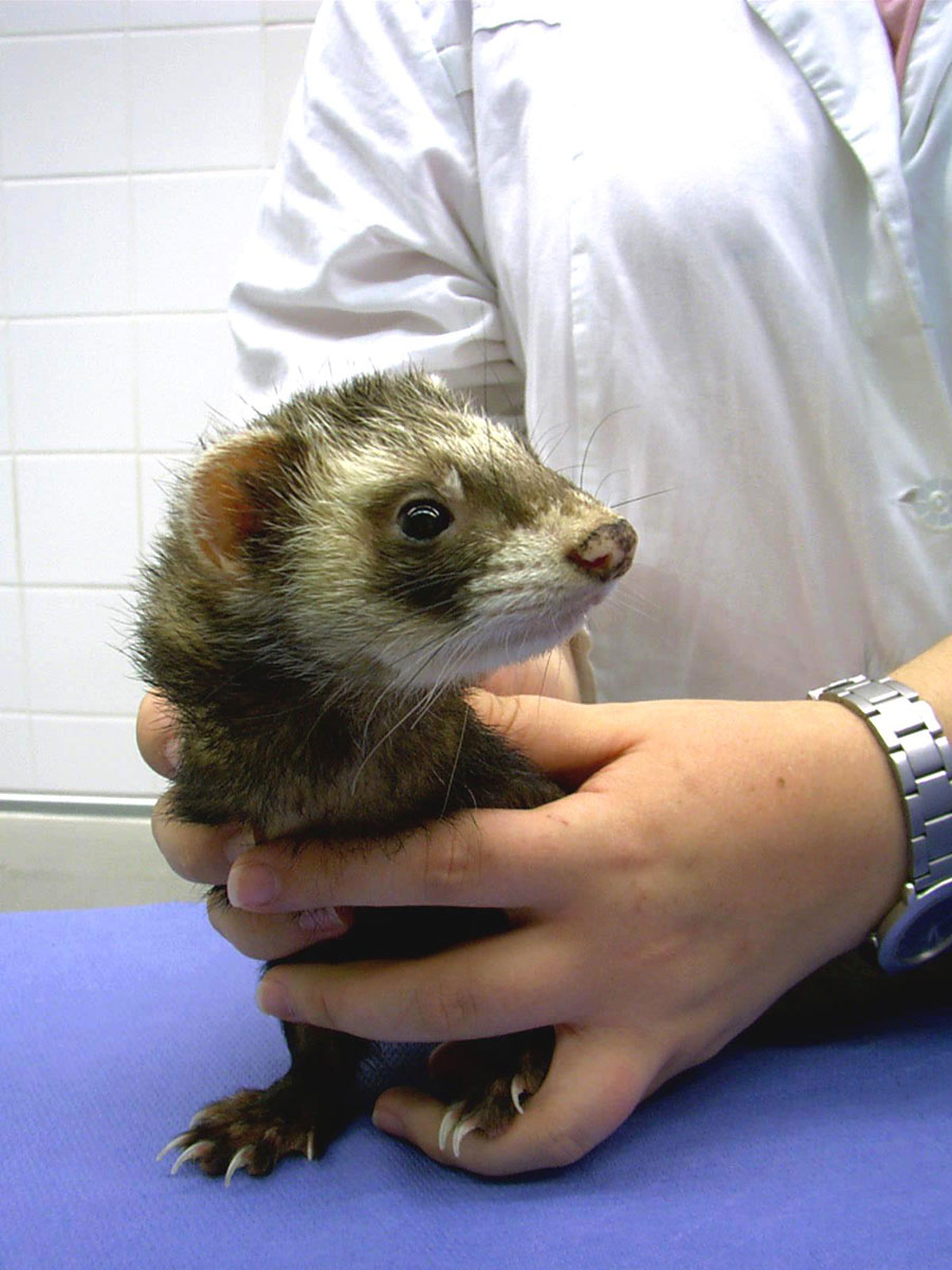
Fig. 1: Ferret during clinical examination.
Image source: Dr. Jutta Hein
Severe acute respiratory syndrome Coronavirus (SARS-CoV-1 & SARS-CoV-2)
SARS-CoV-1 was documented in November 2002, and SARS CoV-2 was reported in December 2019 affecting humans who had contact with infected animals in Chinese markets. Phylogenetic analyses showed great similarities between the two viruses and coronaviruses in cats and bats.
Table 1: Alpha- and beta-coronaviruses relevant to small mammals as well as human medicine
| Species | Alphacoronaviruses | Betacoronaviruses |
| Ferret | Enteric coronavirus (FrECoV), Systemic coronavirus (FrSCoV) |
SARS-CoV-1, SARS-CoV-2 |
| Mink | Mink coronavirus 1 (MCoV) | SARS-CoV-1, SARS-CoV-2 |
| Hamster | – | SARS-CoV-1, SARS-CoV-2 |
| Mouse | – | Mouse hepatitis virus (MHV) |
| Rat | – | Sialodacryoadenitits virus (SDAV) |
| Rabbit | Rabbit Enteric Coronavirus (RECV), Pleural Effusion Disease Virus (PEDV) (not yet assigned) | |
| Human | Human coronaviruses (e. g. HCoV-229E, HCoV-NL63) | Severe Acute Respiratory Syndrome-related Coronavirus (SARS- CoV-1, SARS-CoV-2), Middle East Respiratory Syndrome Corona- virus (MERS-CoV), Seasonal human coronaviruses (HCoV-OC43, HCoV-HKU1) |
Source: Laboklin
They have been assigned to the Betacoronaviruses, but the exact origin and possible intermediate carriers are still unclear. However, a zoonotic origin is most likely. SARS-CoV bind to ACE-2 receptors. Primates, cats, ferrets, syrian golden hamsters and rabbits show a similar receptor distribution to humans and are therefore used as models. With the exception of older animals, ferrets and hamsters showed only mild symptoms in infection trials of SARSCoV-1 and SARS-CoV-2. For SARS-CoV-1, young mice, guinea pigs and rats were also used as models. However, despite virus replication in the tissue, they developed no or only very mild symptoms. For SARS-CoV-2, only transgenic mice with adapted receptors seem to be susceptible.
Mink are particularly susceptible to natural infection with SARS CoV-2. Mink farms in the USA, the Netherlands, Denmark and Spain have experienced major employee-introduced SARS CoV-2 outbreaks with deaths due to severe pneumonia. Proven retransmissions from minks to humans led to the culling of countless mink herds and temporary breeding bans in some countries. Ferrets appear to be much less susceptible. Natural infections with SARS-CoV-2 have been detected in asymptomatic pet ferrets in Spain (8.7 % of 71 animals by PCR, 1.57 % of 127 animals serologically) and in one case in Slovenia.
SARS-CoV-1 and -2 have also been identified in free-living rats in China and New York.
Transmission was most likely through humans via contaminated surfaces as well as sewers. There is no evidence so far that rats led to the spread of the virus. Natural infections with SARS-CoV with clinical symptoms are therefore also possible in small mammals (especially ferrets, rats, hamsters), but rare. SARS-CoV-1 has not played a role since 2004.
Infections with SARS-CoV-2 occur mainly through close contact with infected people in the same household. The incubation period is usually two days. Small mammals are asymptomatic or show mild symptoms (increased temperature, reduced activity, diarrhoea, especially ferrets and hamsters: cough, rhinitis, tracheitis, more pronounced in older animals). Diagnosis is made, as in humans, by PCR from throat swabs. Serological detection of antibodies is possible from two weeks after infection. Antiviral therapy is not available. The animals usually recover within two weeks. Prophylactically, infected people should also observe strict hygiene measures with their pets to avoid infection. So far, there are no reports on infections of humans by their pets, which is probably due to the low virus excretion in animals.
Enteral (FrECoV) and systemic coronavirus of ferrets (FrSCoV)
Epizootic catarrhal enteritis (ECE, synonym «green slime disease») was first described in ferrets in the USA in 1993. The disease is caused by the ferret enteric coronavirus (FrECoV). ECE typically manifests itself as a mucoid green, foul-smelling diarrhoea.
However, initial symptoms can be quite non-specific (lethargy, anorexia and vomiting). The virus is excreted via faeces and saliva. The morbidity is 100 %, but the mortality is quite low at less than 5 %. Older ferrets in particular become severely ill and may die; young ferrets show only mild forms or are subclinically infected (Figure 1). Young animals thus represent a possible reservoir of the pathogen.
The systemic coronavirus of ferrets (FrSCoV) leads to a disease that was initially called «FIP-like disease» because it has many similarities to FIP (feline infectious peritonitis) in cats. It was first recorded in Spain and the USA in 2006. Affected animals first show non-specific symptoms (diarrhoea, anorexia, weight loss, vomiting, sometimes fever). Further symptoms depend on the organs affected. In the case of CNS involvement, central nervous disorders occur (especially weakness/paralysis of the hind legs). Lymph nodes are often enlarged.
The mesenteric lymph nodes in particular are clearly palpable.
Splenomegaly and renomegaly are often present. Pathology shows (pyo-)granulomatous inflammation in the mesentery, peritoneum and affected organs. However, serous effusions such as in FIP are very rare. Changes in blood parameters are very variable (non-regenerative anaemia, hypergammaglobulinaemia, hypalbuminemia, thrombocytopenia). Young animals (< 2 years) are most susceptible.
FrSCoV could also be detected in asymptomatic animals. The onset of systemic disease after infection with FrSCoV is probably caused by a multifactorial process.
The case of clinical manifestation, the course is always progressive, most animals die after a few months or are euthanised.
The seroprevalences of coronaviruses in ferrets in the USA, Japan, the Netherlands and Switzerland range from 32 – 89 % (so far no differentiation between FrECoV and FrSCoV possible). Pathogen detection by PCR from faecal samples showed prevalences of over 60 %. FrECoV was detected more frequently than FrSCoV. Mixed infections have been described. Often, no correlation between pathogen detection and clinic could be established. Despite high prevalences, both disease patterns are seen less and less. To date, there are no prevalence studies from Germany.
Diagnosis is made by direct pathogen detection using PCR. Faecal samples or rectal swabs are suitable for animals with diarrhoea and asymptomatic animals for the identification of carriers, lymph node biopsies or tissue samples for systemically ill animals. Laboklin offers PCR covering the enteric and systemic coronaviruses. The detection of antibodies by ELISA is described, but it is not possible to distinguish between FrECoV and FrSCoV. Since the titre level does not correlate with the symptoms, serology is unsuitable for diagnostics.
Treatment is symptomatic. Antiemetics, antidiarrhoeal agents, infusions, broad-spectrum antibiotics, easy-to-digest food and gastrointestinal protective agents are suitable for ECE. For systemic coronavirus infections, cortisone increase appetite. Doxycycline can help reduce secondary infections and has additional anti-inflammatory effects. Vitamins (vitamin B), minerals and antioxidants can be given to support the immune system. Immunotherapies, derived from the cat’s FIP therapy, can be tried. Prophylactically, the focus is on strict hygiene measures as well as testing of animals that are newly admitted to a herd.
Other coronaviruses in laboratory animals
Other species-specific coronaviruses have been described mainly in laboratory animals. It is unclear as to what role they play in domestic animal populations, as testing for these pathogens is rarely performed.
The mouse hepatitis virus (MHV) leads to enteritis, hepatitis, respiratory diseases or demyelinating encephalomyelitis in mice, depending on the virus strain, but can also be completely absent. In human medicine, it is particularly interesting for research into hepatitis, multiple sclerosis and SARS, for example.
Sialodacryoadenitits virus (SDAV, rat coronavirus) causes respiratory diseases (rhinitis, tracheobronchitis, pneumonia) in rats associated with inflammation of the salivary and lacrimal glands.
Rabbits can also be infected with coronaviruses. Pleural effusion disease virus (PEDV) leads to pleural effusions, cardiomyopathy, mesenteric lymphadenopathy and multifocal necrosis of various organs. The pathogen has so far only been found in laboratory animals in North America and Europe. Rabbit enteric coronavirus (RECV) causes enteritis, especially in young rabbits. Apart from laboratory rabbits, it it also plays a role in rabbit farms in Europe and the USA.
In North America, the seroprevalence is between 3 – 40 %.
Conclusion
FrECoV and FrSCoV are particularly relevant to the veterinarian and should be considered in ferrets as a differential diagnosis for diarrhoea and/or systemic disease. SARS-CoV-2 infections should be taken into consideration when respiratory infections suddenly occur in ferrets, rats or golden hamsters in households with currently Corona-infected individuals, as the infection occurs via humans.
Dr. Ekaterina Salzmann, Dr. Jutta Hein, Jana Liebscher


