Infections with Echinococcus are some of the most important zoonoses caused by cestodes. In Germany, the disease is notifiable in humans and some animal species, especially dogs. Currently, there are eight known species in the cestode genus Echinococcus:
The fox tapeworm (Echinococcus (E.) multilocularis) is exclusively present in the Northern Hemisphere. The core endemic areas include Southern Germany, Switzerland, Central- and Eastern France as well as Western Austria. The dog tapeworm (E. granulosus sensu lato (s.l.)) is distributed worldwide. In Europe, it can be found mainly in the East and South as well as along the Mediterranean Coast. This cestode is rare in Germany. E. granulosus s.l. is a species complex, which currently consists of five different species with different morphological, biological and genetical characteristics, these are E. granulosus sensu stricto (genotypes G1 – G3), E. felidis, E. equinus, E. ortleppi and E. canadensis (genotypes G6/G7, G8 and G10). The species E. vogeli and E. oligarthrus can only be found in Central and South America.
As definite hosts, dogs pose a significant risk of transmission of E. multilocularis and E. granulosus s.l. to humans. In fact, keeping dogs as pets is listed as one of the most important risk factors for humans to become infected with these parasites. The following article will cover the basics of life cycle, prevalence as well as diagnostic methods of Echinococcus and therefore provide a foundation from which to estimate the potential risk of infection posed by dogs.
Life cycle
Echinococcus cestodes present with an indirect life cycle featuring definite and intermediate hosts. The sexually active, mature tapeworms parasitize in the small intestine of carnivores. Unlike other cestodes, these worms only show a length of a few millimetres and consist of a scolex (head) and 2-6 proglottids (table 1).
The proglottids containing the eggs are shed from the rest of the worm. In contrast to the proglottids of other tapeworms, the ones of Echinococcus often already break up in the intestine and release the instantly infectious eggs into the faeces.
The eggs contain an oncosphaere (1st larval stage), which will be (most commonly) ingested orally by the intermediate hosts. In the intermediate hosts, the oncosphaere first penetrates the intestinal wall and then migrates, via the bloodstream, to the internal organs, especially the liver. There, the larvae develop into metacestodes (2nd larval stage), which usually take the shape of hydatid cysts.
As soon as metacestodes have developed protoscolices (immature cestode scolices), the larvae are fertile. The life cycle is concluded when the definite host ingests infected organs of the intermediate host. In the small intestine of the definite host, the scolex everts and attaches itself to the intestinal wall and the worm matures into an egg-producing adult. Beside the intermediate hosts, accidental / aberrant hosts, which are not part of the natural life cycle, can also become infected by ingesting the eggs. Metacestodes also develop in the organs of accidental hosts.
- Source: Dr. Michaela Gentil
-
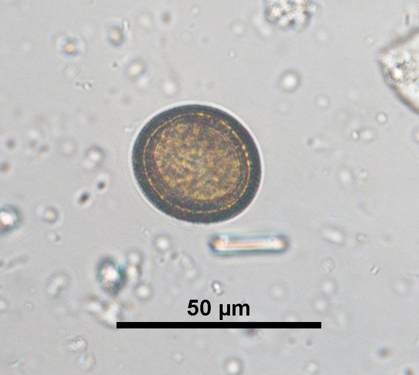
Figure 1: Taeniidae cestode egg in a faecal sample of a domestic dog
Source: Laboklin
-
Figure 2: Liver of a domestic dog with alveolar echinococcosis, fixed in formalin
Source: Laboklin
-
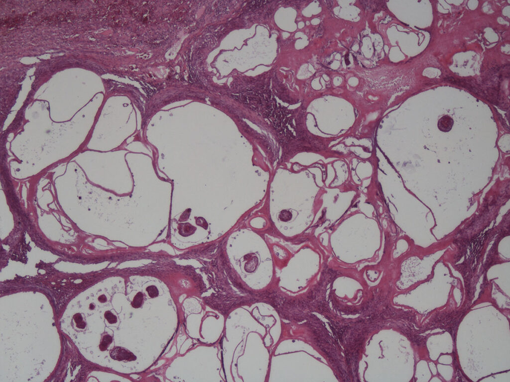
Figure 3: Histology of a cystic liver lesion, featuring cross sections of protoscolices, HE stain, 40x magnification
Source: Laboklin
Table 1: Important characteristics of E. multilocularis and E. granulosus s.l.
| Species | Length of the adult stages | Number of proglottids | Number of eggs | Prepatency | Patency |
| E. multilocularis | 1.2 – 4.5 mm | 5 (2 – 6) | Up to 200 | 28 days | Several months |
| E. granulosus s.l. | 2 – 7 mm | 3 (2 – 6) | Up to 1500 | 45 days | Several months |
The life cycle of Echinococcus requires different animal species as hosts (table 2). Definite hosts are mainly canids, very rarely, felids are affected. The host species has an impact on larval development and the fecundity of the adult worms: in felids, the parasites develop much slower and produce, if at all, only low numbers of eggs.
Table 2: Definite, intermediate and accidental / aberrant hosts E. multilocularis and E. granulosus s.l.
| Species | Definite hosts | Intermediate hosts | Aberrant hosts |
| E. multilocularis | Mainly foxes (red fox, polar fox), racoons and racoon dogs, also wolves, domestic dogs, (cats) and other carnivores | Small mammals (especially rodents) | Humans, primates, domestic dogs, (house pig and wild boar), and others |
| E. granulosus s.l. | Domestic dog, other canids (e.g. wolve, coyote), possibly felids | Mainly sheep, other ruminants, pigs, horses and others | Humans, various other mammals |
Clinical symptoms
In the definite hosts, infections with adults of Echinococcus are inapparent without any symptoms in most cases, even in severe infestations. The low pathogenic adult cestodes attach with their scolices to the intestinal mucosa, any localized lesions are usually inconsequential. However, definite hosts pose a risk of infection towards humans. In domestic dogs, which have been tested positive or are under suspicion to be infected, it is imperative to start treatment immediately, while adhering to strict hygiene and safety procedures. For more information, as well as for possible preventive measures, please see the ESCCAP-Guidelines.
After ingesting Echinococcus eggs, metacestodes develop in the internal organs of intermediate and aberrant hosts (especially humans), where they show a high pathogenicity and can cause severe disease.
Echinococcus granulosus sensu lato
In the intermediate and aberrant host, the clinical picture is known as cystic echinococcosis. A hydatid cyst is formed, which can reach a diameter of a few decimetres. The cystic wall consists of several layers, the outer layer is formed by host connective tissue, followed by a laminated membrane and a germinal epithelium. From this epithelium, vacuoles form, so-called brood capsules, in which the protoscolices develop. Clinical problems arise mainly from pressure building up towards the surrounding tissues and organs when the cysts increase in volume. It can take several years for the disease to become apparent.
Echinococcus multilocularis
The small fox tapeworm causes alveolar echinococcosis in intermediate and aberrant hosts, which is an exceptionally severe disease with a high mortality rate if left untreated. In contrast to cystic echinococcosis, the metacestode does not form an enclosed cyst, but infiltrates the surrounding tissue and grows tumor-like, constituting multiple vesicles. In most cases, only the liver is affected. The germinal epithelium sprouts into the surrounding tissue, where multiple protoscolices develop. In human cases, it can take up to 10-15 years until the disease becomes apparent. However, there is also a large percentage of aborted infections, in which the immune system manages to terminate parasite development and therefore stop the disease right at the start.
The domestic dog plays a special role in the life cycle of E. multilocularis, since dogs can be definite as well as aberrant hosts. Definite hosts become infected by ingesting small mammals (like rodents) which contain the metacestodes. Therefore, dogs known for hunting and eating rodents are particularly at risk.
Occasionally, domestic dogs become aberrant hosts by ingesting the eggs of E. multilocularis and which manifests as alveolar echinococcosis as described above. In these cases, infectious eggs were present in the environment, the dog practiced coprophagia or the dog was primarily infected itself intestinally. Eggs shed by faeces can stick to the fur coat and from there, can be transmitted to the dog and its owners.
In dogs suffering from alveolar echinococcosis as aberrant hosts, one should always check for a concurrent intestinal infection. If positive, then there is an actual risk of infection for humans who came into contact with the dog (owners, staff at the veterinary practice). In the case of a negative result, it cannot be ruled out that an infection took place at an earlier date. Therefore, dog owners should be made aware of the possibility of exposure as well as available serological diagnostic methods.
The most promising therapy for canine alveolar echinococcosis is the complete surgical removal of the metacestodes, taking care to leave a safety margin to healthy tissue, combined, if necessary, with a benzimidazole treatment. In many cases, this disease will be diagnosed at a late stage of infection and therefore, it will be too late for a surgical intervention due to complete infestation of liver / lung and / or extrahepatic metastases.
Diagnostic methods
Diagnosing the definite host
Proglottids only exhibit a length of a few millimetres and often break up during the intestinal passage.
Therefore, they cannot be identified with the naked eye.
Eggs of the family Taeniidae, which encompasses both the genera Taenia and Echinococcus, are round, possess a thick shell with a radially striped, brown coloured embryopore (figure 1). Their diameter is between 30 to 40 µm and they contain the first larval stage. Eggs can be found coproscopically using flotation and sedimentation methods, however, eggs of different members of the family cannot be differentiated morphologically. To distinguish Echinococcus from Taenia eggs, which have a low zoonotic significance, a variety of faecal antigen tests as well as molecular methods are available from specialized diagnostic laboratories, e.g. PCR tests can be used to identify different species.
False negative results are possible in some cases, since the proglottids / eggs are shed intermittently, few eggs will be present with a low worm burden and during the prepatency, no eggs are shed since the worms have not matured yet.
Adult worms are mainly found during post-mortem examinations. The species can be identified using differed characteristics like morphology of the proglottids and reproductive tract, as well as number, form and size of the hooks surrounding the scolex. There is only a weak correlation between intestinal infections and the presence of Echinococcus antibodies in the serum. Serological tests in definite hosts are therefore of low clinical value, especially since these tests cannot discern between intestinal and alveolar echinococcosis in domestic dogs.
Diagnosing the intermediate and aberrant hosts
Cysts in affected organs can be detected using diagnostic imaging techniques. A macroscopic detection of tissue lesions, especially in the liver, can be attempted with laparotomy in living animals and during a post mortem examination (figure 2). Histology of cystic tissue (figure 3) and / or PCR tests can help to confirm the diagnosis.
Prevalence of Echinococcus in dogs in Germany
During a study on faeces samples of dogs in Europe (n = 21588), Dyachenko et al. 2008 found a 0.25% prevalence of Taeniidae eggs. In 43 of the 53 dogs positive for Taeniidae eggs, a species differentiation identified an infection with E. multilocularis (81%).
Our own sample material yielded similar results, an evaluation of European canine faeces samples (n=60615), using flotation and sedimentation, revealed a Taeniidae prevalence of 0.16%. Following their detection, 35 of these Taeniidae positive samples were submitted to a Echinococcus PCR test. In 22 of these samples (62.9%), E. multilocularis was detected.
Therefore, a detection of Taeniid eggs in dog faeces should always be treated as a potential infection with E. multilocularis, for which further diagnostic differentiation is imperative due to the particular zoonotic potential.
In contrast, E. granulosus s.l. could not be detected in European dogs, neither by Dyachenko et al. (2008), nor in our own cumulative test results.
Despite this, intestinal infections, especially in dogs imported or travelled from endemic regions, should always be taken into account as a differential diagnosis.
Due to the increase in the fox population in the past decades, as well as the expansion of foxes into the urban environment, the infective pressure has risen and with it the number of human cases. There is, however, no data available concerning alveolar Echinococcosis in domestic dogs, neither for Germany nor in a global context.
Conclusion
As the definite hosts for E. multilocularis, domestic dogs pose a relevant infectious risk for humans in Central Europe. In addition, dogs can become aberrant hosts themselves and present with clinically apparent echinococcosis. Therefore, it is imperative to prevent infections of domestic dogs. The prevalence of infections with cestodes of the family Taeniidae in dogs from Europe is low, however, the proportion of E. multilocularis positives is high. Since cestode eggs of the family Taeniidae cannot be distinguished microscopically, infections with Echinococcus have to be identified using specific diagnostic methods.
Dr. Michaela Gentil
Service portfolio
- Parasitological examination (flotation / sedimentation) of faecal samples
- Specific PCR-test for the detection of Echinococcus multilocularis and Echinococcus granulosus in faeces, cyst material and tissue
- Detection of Echinococcus antibodies in serum using ELISA


