The Laboklin Expert Panel has now become a well-established institution. Three to four times a year, it addresses a broad range of clinically relevant questions. For the topic of Cushing’s syndrome, specialists in endocrinology, pharmacology, and surgery shared their expertise, drawing on both current clinical experience and the latest scientific evidence.
The experts participating in the panel were:
Prof. Dr Wolfgang Bäumer, Dipl. ECVPT, Director of the Institute of Pharmacology and Toxicology, Faculty of Veterinary Medicine, Free University of Germany; Prof. Dr Nadja Sieber-Ruckstuhl, Dipl. ACVIM and ECVIM-CA, Head of Endocrinology, Small Animal Clinic, University of Switzerland; PD Dr Astrid Wehner, Dipl. ECVIM-CA, Senior Lecturer in Internal Medicine with a focus on Endocrinology, LMU Germany; PD Dr Florian Zeugswetter, Head of the Endocrinology Department, University Small Animal Clinic, Austria; Dr Pieter Nelissen, Dipl. ECVS, RCVS Specialist, Managing Director and Chief Surgeon, Frontier Small Animal Specialists, Germany.
The introduction addresses the current nomenclature of Cushing’s syndrome. PD Dr Florian Zeugswetter explains that the European Society of Veterinary Endocrinology (ESVE), within the framework of the “ALIVE” project, has agreed on a standardised terminology. Disorders caused by an excess of glucocorticoid-active substances are therefore referred to as Cushing’s syndrome (CS). A distinction is made between iatrogenic forms and naturally occurring CS.
Naturally occurring CS can be further classified into ACTH-dependent and ACTH-independent forms.
ACTH-dependent variants include the classic pituitary-dependent CS, whereas ACTH-independent forms result from autonomously hormone-producing adrenocortical tumours. In addition, special forms exist, such as subclinical (formerly “atypical”) CS, in which clinically typical signs are present, but established functional tests do not allow a definitive diagnosis.
This systematisation aims both to facilitate scientific communication and to improve clinical classification.
The next part of the panel focuses on the clinical symptomatology. Prof. Dr Nadja Sieber-Ruckstuhl emphasises that the majority of dogs with CS exhibit the classic signs of polyuria and polydipsia (PU/PD) as well as polyphagia. PU/PD is reported in over 80 % of cases, while increased food intake occurs in more than 50 %. She notes, however, that a certain proportion of patients do not show a clear manifestation of these symptoms. This may in part be attributed to increased awareness among dog owners and veterinarians of early, subtle clinical signs, resulting in affected animals being presented at an earlier stage of the disease. During the general clinical examination, notable findings include a distended but soft abdomen, thin and rather dry skin, abnormal fat distribution (e.g., central obesity), and muscle atrophy.
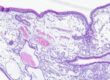
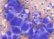
The participants asked about calcinosis cutis, how it is related to CS, and how it can be treated. PD Dr Florian Zeugswetter explains that it involves calcium deposits in the skin, which predominantly occur in the neck and back regions, but can also appear in the inguinal area or on the extremities. Its occurrence is described almost exclusively in association with glucocorticoid exposure, making calcinosis cutis nearly pathognomonic for CS. Certain breeds, such as Staffordshire Bull Terriers, Pitbull Terriers, and Rottweilers, show a particular predisposition. Clinically, these skin changes are often striking and may vary in severity. Prof. Dr Wolfgang Bäumer notes that the best therapeutic approach is consistent management of Cushing’s syndrome. Supportive measures, such as keratolytic shampoos or DMSO solutions, are discussed, although the evidence supporting their use is limited.
When asked about the prevalence of systemic hypertension in dogs with CS, PD Dr Astrid Wehner emphasises that approximately 80 % of CS patients are affected. Close monitoring is recommended for a systolic blood pressure of 160 mmHg or higher, while values above 180 mmHg carry a high risk of target organ damage, particularly at the renal and cardiovascular level.
Therefore, blood pressure measurement should be performed in all patients with CS.
PD Dr Astrid Wehner also provides a brief digression on proteinuria. Almost every second dog with CS exhibits proteinuria, with the urine protein-to-creatinine ratio (U-P/C) typically ranging from 1 to 3. However, more severe proteinuria with considerably higher U-P/C ratios is also possible.
The exact cause is unclear. In some patients, concomitant systemic hypertension is certainly contributory. PD Dr Wehner points out that study data demonstrate both glomerular sclerosis and tubular lesions. Nevertheless, most patients do not develop azotaemia despite these changes, and proteinuria does not appear to have prognostic significance. It often regresses under successful therapy, although not in all cases. PD Dr Wehner agrees with PD Dr Zeugswetter and Prof. Dr Sieber-Ruckstuhl that CS-associated proteinuria in dogs does not require separate treatment.
Cats can also develop Cushing’s syndrome, although it is significantly less common than in dogs. Dr Astrid Wehner addresses the differences compared with dogs, explaining that feline CS is frequently associated with diabetes mellitus. While this can also occur in dogs, it is not seen to the same extent: up to 80 % of cats with CS are diabetic.
Clinically, polyuria and polydipsia associated with often poorly controlled diabetes mellitus predominate. However, in contrast to dogs, these symptoms are considerably less pronounced in cats without diabetes mellitus. As in dogs, increased food intake is a typical clinical feature of CS. Affected cats lose weight, accompanied by marked muscle wasting, which is also a common finding in dogs.
Cats, like dogs, often present with a characteristic, pendulous, distended abdomen. The skin is thin, dry, and scaly, as is the case in dogs with CS, so the abdominal veins are particularly visible in both species. In cats, this effect is so pronounced that it leads to marked skin fragility. Consequently, poorly healing wounds are common, and even relatively minor mechanical stress can lead to extensive skin tears. The course of the disease in cats is usually insidious, which complicates early detection. Dr Pieter Nelissen notes that adrenal tumours are somewhat less common in cats than in dogs but still represent an important differential diagnosis. Adrenal cortical tumours in cats may produce cortisol as well as aldosterone or sex hormones, either concomitantly or instead of cortisol. Symptoms can be very similar, but diagnosis is more challenging. Prof. Dr Nadja Sieber-Ruckstuhl explains that altered sexual behaviour, such as sudden urine marking in neutered male cats or signs of oestrus in female cats, can sometimes provide a clue. During the general examination, in suspected cases, castrated male cats should be checked for penile spines, which are normally present only in intact animals.
The discussion then briefly addresses iatrogenic Cushing’s syndrome. It occurs relatively frequently in dogs and less often in cats. The question arises as to the dose and type of administered glucocorticoids at which it can be expected. Prof.
Dr Nadja Sieber-Ruckstuhl emphasises that this cannot be answered universally. Iatrogenic CS can be triggered by very low doses of glucocorticoids, particularly when administered over a prolonged period. There is a high degree of individual sensitivity, with large dogs often being particularly susceptible. The test of choice in cases where iatrogenic CS needs to be distinguished from naturally occurring CS is the ACTH stimulation test. In iatrogenic CS, it yields a result that would typically be expected for hypoadrenocorticism (no stimulation or borderline stimulation).
Another central topic is the diagnosis of CS. The discussion begins with the challenge posed by prior glucocorticoid treatment. Prof. Dr Nadja Sieber-Ruckstuhl explains that there are no validated guidelines in the literature regarding the optimal interval between glucocorticoid administration and a functional test. This interval depends on the specific preparation, dose, duration of administration, and individual sensitivity. In some patients, normal stimulation can be observed one week after withdrawal of exogenous glucocorticoids, while in others it may take months before there is no residual effect. As a general guideline, 6–8 weeks are often suggested. Prof. Dr Wolfgang Bäumer adds that numerous medications can influence cortisol levels: for example, butorphanol increases cortisol concentrations, whereas substances such as trazodone, lokivetmab, and bedinvetmab can reduce them—particularly by lowering stress and pain. This necessitates especially careful interpretation of test results.
The “ALIVE” group of the ESVE explicitly advises against the use of hormone tests for in-house diagnostics.
PD Dr Florian Zeugswetter agrees with the other experts that a single basal cortisol measurement is unsuitable for diagnostic purposes. In dogs with CS, cortisol overexposure typically results from an increased frequency of secretory peaks. A randomly obtained blood sample may therefore coincide with either a peak or a trough of cortisol secretion. Consequently, it is not possible to reliably distinguish affected from unaffected patients, regardless of whether the measured value is high or low.
The urine cortisol-to-creatinine ratio (UCC) can provide an initial orientation but should always be supplemented with functional tests. Prof. Dr Nadja Sieber-Ruckstuhl recommends analysing at least three individual samples, as pooled urine samples may distort the results. Urine should be collected at home by the owner. Following a veterinary visit, at least two days should elapse before starting the collection, as stress induced by the visit could result in false positive results.
PD Dr Florian Zeugswetter provides the participants with a more detailed discussion of the low-dose dexamethasone suppression test (LDDST), which is generally regarded as the test of choice for confirming the diagnosis of CS. However, the LDDST can be influenced by situations that increase cortisol secretion, such as emotional or disease-related stress. A positive result therefore confirms CS only in the context of compatible clinical signs and the exclusion of other diseases.
Following dexamethasone administration, physiological feedback suppresses endogenous cortisol production. A reduction of serum cortisol concentration below a defined cut-off is expected. In most patients with Cushing’s syndrome, this suppression is absent. Interpretation of the test is primarily based on the 8-hour value. For a definitive assessment, reduced suppression should also be evident in the intermediate time point (typically at 4 hours). Classically, the test is considered negative (i.e. CS is not present) if the 8-hour value is below the established cut-off, regardless of the intermediate value obtained at 3–4 hours.
However, a negative result is not necessarily conclusive; therefore, in cases of persistent clinical suspicion, repeat testing or the use of alternative diagnostic methods may be required.In cats, the LDDST is also considered the method of choice, although the dexamethasone dose must be adjusted to 0.1 mg/kg—ten times higher than in dogs.
Further questions from the audience concern the practical execution of the LDDST. One topic is the concentration of dexamethasone preparations. Some confusion arises from certain literature sources discussing the conversion of effective concentrations. Prof. Dr Wolfgang Bäumer explicitly refers to the concentration stated on the preparation itself. In German-speaking countries, this is always provided (“corresponds to x mg dexamethasone”). Additionally, participants asked how dexamethasone can be practically diluted for very small patients. PD Dr Florian Zeugswetter provides guidance: adding 1 ml of a 2 % dexamethasone preparation (2 mg/ml) to 9 ml of 0.9 % NaCl (mixed well) results in a final concentration of 0.2 mg/ml, which allows for easier dosing. For a dexamethasone concentration of 4 mg/ml, 0.5 ml of dexamethasone is added to 9.5 ml of 0.9 % NaCl to achieve the same final concentration of 0.2 mg/ml.
With regard to therapy, medical treatment with trilostane plays a central role. This drug inhibits cortisol synthesis through reversible enzyme blockade. Prof. Dr Wolfgang Bäumer notes that, despite its reversibility, irreversible adrenal cortical necrosis may occur in rare cases, leading to permanent hypoadrenocorticism. Concerning dosage, twice-daily administration of lower amounts is preferable to a single high daily dose. This is particularly beneficial in large-breed dogs, where it significantly reduces the risk of adverse effects. PD Dr Astrid Wehner advises caution in patients with chronic kidney disease. Reducing cortisol can lower the glomerular filtration rate and may consequently worsen the course of renal disease. Dr Pieter Nelissen adds information on surgical options: in cases of adrenal tumours, adrenalectomy is the treatment of choice, as approximately 50 % of these tumours are carcinomas. The complication rate of such surgery has been markedly reduced in recent years due to advances in surgical techniques and anaesthesia. Even when vascular invasion is present, surgical removal may still be possible. If a patient is already receiving trilostane, the medication should be discontinued 24–48 hours prior to surgery. The remaining contralateral adrenal gland is typically atrophied and requires time to regain function.
Postoperatively, a low dose of corticosteroids should therefore be administered and tapered over 4–6 weeks.
Monitoring was also discussed in detail. PD Dr Florian Zeugswetter favours pre-pill cortisol measurement as a cost-effective option, provided the patient’s clinical condition is stable and there is no suspicion of trilostane overdose. Blood sampling is carried out immediately before the next scheduled dose of trilostane. Prof. Dr Sieber-Ruckstuhl recommends double sampling at a one-hour interval to account for the effects of random peaks and stress responses. In this approach, one cortisol measurement is taken at the time when the trilostane dose would normally be administered, and a second measurement one hour later. The trilostane is only given after the second blood draw on that day. This procedure allows stress-induced high or incidentally low cortisol concentrations to be “balanced out”, potentially leading to a more accurate assessment of the patient. PD Dr Wehner reminds us that reduced appetite or decreased vitality may be early warning signs of trilostane overdose. The ACTH stimulation test offers the highest level of safety when assessing potential overdosing and should always be used when the patient shows clinical abnormalities. She also emphasises that the risk of therapy-induced hypoadrenocorticism increases with treatment duration. Close, lifelong monitoring using ACTH stimulation or pre-pill cortisol measurements are therefore indispensable.
Dr. Jennifer von Luckner