The senses of hearing and balance are located in the ear. Anatomically, the ear has three compartments: the outer, middle and inner ear. Inflammation of any part of the ear is called otitis. Otitis externa is the most frequent and one of the most common problems in dermatology consultations. An incorrect approach to otitis leads to failure and compromises the viability of the ear and the quality of life of the patient and owner. Otitis externa has a multifactorial origin, with primary causes, predisposing and perpetuating factors involved in the inflammation of the external ear canal (EEC), creating a suitable environment for the proliferation of micro-organisms (secondary cause). Successful treatment and resolution of otitis externa depend on the clinician’s expertise in identifying and treating every factor involved.
1. Primary causes are the processes capable of producing otitis externa alone. They must be identified and treated to avoid chronification and recurrence of otitis externa. The most common primary causes are allergic diseases, foreign bodies, ectoparasites (Otodectes cynotis, Demodex), masses (polyps, neoplasms), endocrinopathies, and less frequently contact dermatitis, irritant dermatitis, autoimmune diseases or drug reactions.
2. Predisposing factors are conditions that may favour the development of otitis, and these include:
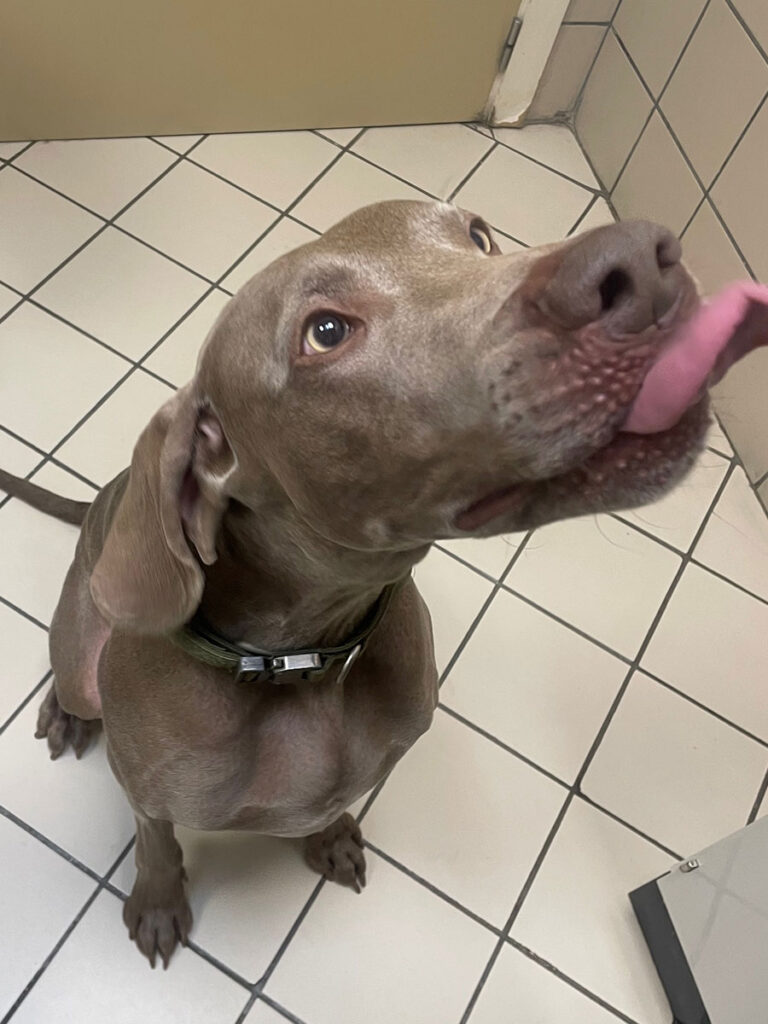
- Anatomical characteristics, e.g. breed-associated stenosis of ear canal, drooping ears (Fig. 1)
- Using traumatic techniques such ascleaning with cotton swabs or hairremoval from the ear canaly
- Moisture in ear canals, e.g. animals who swim
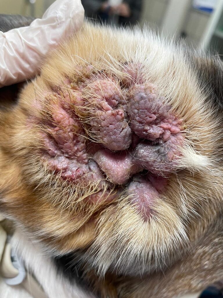
3. Perpetuating factors are progressivepathological anatomical changes (oedema,epithelial hyperplasia, hyperplasia ofceruminous glands, stenosis, fibrosis ormineralisation of the EEC, perforation ofthe tympanic membrane, otitis media),consequence of the chronicity of theinflammation, which hinders the resolution ofotitis externa and perpetuates it (Fig. 2).
4. Secondary causes represent the infectiouscomplication bacteria or yeasts. Primarycauses, predisposing and perpetuating factorsgenerate ideal conditions for colonisation andmultiplication of micro-organisms.
Diagnostic approach to otitis externa
Clinical signs
Shaking or tilting of the head, scratching of the ears or head, presence of exudate, foul odour or pain are common reasons for consultation. Foreign body and mite otitis usually present the most prominent acute clinical signs, mainly because of their acute painful or pruritic condition.
The presence of pain on opening the mouth or neurological signs such as head tilting, Horner’s syndrome, nystagmus, ataxia, loss of balance and walking in circles suggest the existence of otitis media or internal otitis.
Physical examination
Visual examination of the pinnae and the opening of the EEC should assess for the presence of exudate and lesions such as erythema, epidermal hyperplasia, excoriations and erosions /ulcerations. A foul odour may be detected.Palpation of the ear canals, which should be carried out gently, helps determine whether there is pain or pruritus. If an otitis externa is suspected, otoscopy or video-otoscopy and cytology of the exudate should be done.
Otoscopy/video-otoscopy
Otoscopy allows assessment of the integrity or pathological changes of the external ear canal (EEC) and tympanic membrane. Pulling the pinna aligns the vertical and horizontal portions of the EEC and facilitates visualisation of the canal down to the tympanic membrane.
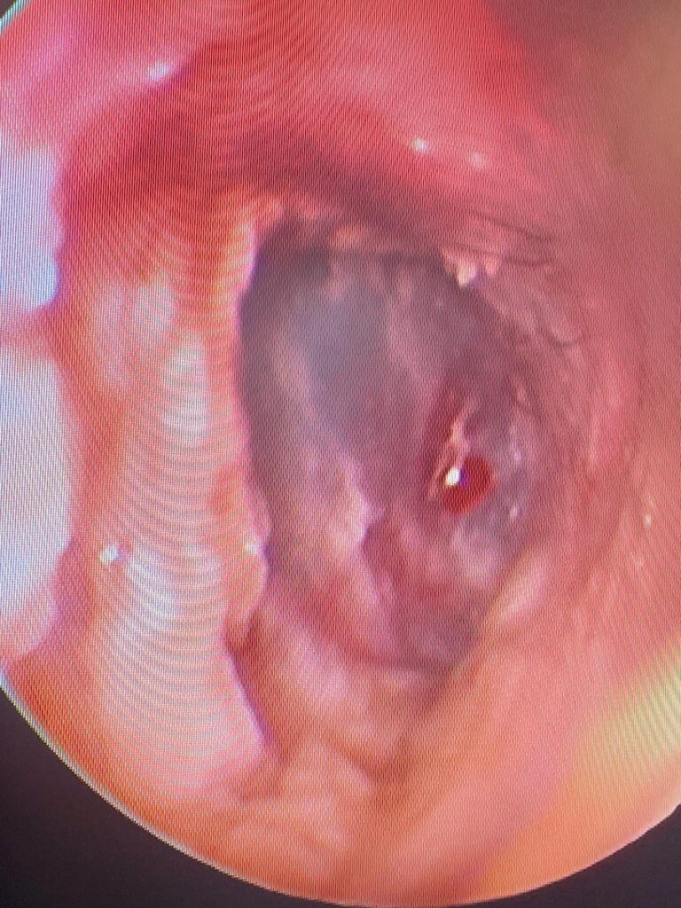
The EEC should be patent and exudate-free in a healthy ear and visualisation of the eardrum should be possible. The exudate, cerumen or cerumenoliths, the inflammation and/or the stenosis of the EEC may prevent visualisation of the tympanic membrane. In the case of otitis, otoscopy allows evaluating the quantity and characteristics of the exudate and the alterations of the EEC: erythema, oedema, hyperplasia, stenosis, ulceration and presence of masses (Fig. 3).
In case of severe pain, ulceration or EEC stenosis, anaesthesia would be required. However, it is recommended to postpone the otoscopy until these conditions are controlled, except in cases of suspected foreign body, as it is essential to facilitate its removal.
-
Fig. 1: Drooping ears are a predisposing factor for otitis externa.
Source: Dr Carmen Lorente
-
Fig. 2: Severe chronic otitis with hyperplasia of the ear skin and complete narrowing of the ear canal opening.
Source: Dr Carmen Lorente
-
Fig. 3: Videotoscopy during deep ear flushing under general anaesthesia: erythema and oedema of the ear canal walls and inflammation of the eardrum are observed.
Source: Dr Carmen Lorente
-
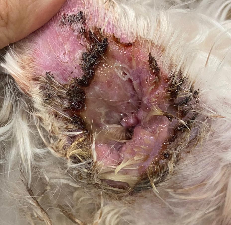
Fig. 4: Purulent otitis: fluid exudate from the external ear canal, forming crusts on contact with air. Cytology showed neutrophilic inflammatory component with rod bacteria.
Source: Dr Carmen Lorente
Microscopic evaluation of otic exudate
Evaluation of otic exudate is used to detect parasites (direct microscopic exam) and infectious agents (cytology).
Direct microscopic exam
A sample of otic exudate obtained with a swab or curette is placed in a drop of oil on a slide. The exudate is dispersed in the oil and a coverslip applied. Under 4x magnification, the presence of Demodex or Otodectes diagnoses the primary cause of otitis.
Cytology
Depending on the type of exudate, otitis are classified as ceruminous, bacterial, yeast, mixed or purulent.
Ceruminous exudate consists of cornified epithelial cells and lipids and may contain a small number of coccoid bacteria and yeasts (up to 5 yeasts and 25 coccoid bacteria per 40X field may be expected). The presence of rod bacteria is always considered pathological.
The proliferation of bacteria and/or Malassezia characterises otitis into coccoid, rod or mixed bacterial; Malassezia or mixed otitis (bacteria and Malassezia).
The presence of inflammatory cells (neutrophils or neutrophils and macrophages) defines purulent otitis (Fig. 4). A purulent exudate may be observed in pemphigus foliaceus and infectious otitis, especially otitis with rod-shaped bacteria.
Bacterial culture and sensitivity
Initially, it is unnecessary since with topical treatment, the antibiotic concentration to which the bacteria are exposed is much higher than that which can be obtained systemically.
When to perform culture and bacterial sensitivity?
- When the antibiotic initially selected doesnot resolve the infectiony
- Chronic or recurrent otitis in which severalantibiotic treatments have been usedy
- Presence of rod-shaped bacteria oncytologyy
- Otitis media
Never use quinolones without justified cause, that is when an antibiogram demonstrate resistance to first-choice antibiotics.
Other diagnostic tests useful in otitis
In cases where middle or inner ear involvement is suspected, imaging techniques should be used. Computerised axial tomography (CAT) allows visualisation of the eardrum, assessment of the contours of the tympanic bulla and detection of the presence of bony proliferations and osteolysis. Magnetic resonance imaging (MRI) differentiates between fluid and soft tissue but does not detect bone changes easily.
Treatment
Failure to treat all the causes and factors invol-ved results in chronic or recurrent otitis externa with proliferative changes that may require aggressive surgical treatment.
It is crucial to control inflammation and its cause. Inflammation and exudates make an ideal environment for the proliferation of bacte-ria and Malassezia. Topically applied corticoste-roids are usually enough to control inflamma-tion, but oral administration may be necessary if severe inflammation occurs. Topical administration is sufficient and more effective if antibiotic or antifungal treatment is necessary. Systemic antibiotics or antifungals are not required in otitis externa unless there are concurrent otitis media.
Thorough cleaning of exudates is crucial. If uncomplicated ceruminous otitis using otic cleansers. If severe otitis with intense microbial growth, rod-shaped bacteria, profuse or purulent exudate, or biofilm formation, performing a thorough cleaning with a video-otoscope and under general inhalation anaesthesia is ideal.
Dr. Carmen Lorente Méndez,
DVM, PhD, DipECVD
Further reading
-
Bischoff MG, Kneller SK. Diagnostic imaging of the canine and feline ear. Vet Clin North Am Small Anim Pract. 2004 Mar;34(2):437-58. doi: 10.1016/j.cvsm.2003.10.013. PMID: 15062618.
-
Gotthelf, L. N. Small animal ear diseases: an illustrated guide. 2nd editon. St. Louis: Elsevier/Saunders;2005
-
Nuttall T, Bensignor E. A pilot study to develop an objective clinical score for canine otitis externa. Vet Dermatol. 2014 Dec;25(6):530-7, e91-2. doi: 10.1111/vde.12163. Epub 2014 Aug 6. PMID: 25130194.
-
O’Neill DG, Volk AV, Soares T, Church DB, Brodbelt DC, Pegram C. Frequency and predisposing factors for canine otitis externa in the UK – a primary veterinary care epidemiological view. Canine Med Genet. 2021 Sep 7;8(1):7. doi: 10.1186/s40575-021-00106-1. PMID: 34488894; PMCID: PMC8422687.


