Enlarged toes are often painful and cause lameness. They are often difficult or impossible to treat using antibiotics.
Amputation of the toe is then the only promising treatment option. It is a common surgery in enlarged toes of dogs and cats and patients usually recover well.
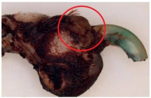
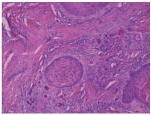
Amputations also offer the opportunity to both treat and diagnose the cause of the lesions, since the clinical presentation of neoplastic and non-neoplastic (Fig.1) lesions can be very similar.
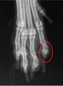
Destruction of the bone in particular can be caused by malignant and benign tumors as well as by inflammation and other non-neoplastic lesions (e.g. hair follicle cysts). Bone destruction is often detected radiographically prior to toe amputation (Fig. 2). On the other hand, there are also cases in which no skeletal abnormalities are seen in which malignant bone destructive tumors are later diagnosed histologically.
Amputated toes should therefore always be examined by histopathology.
There is little data available on histopathological findings in amputated toes in dogs and cats.
The following analysis therefore looks at the histological results of 380 canine and 87 feline toe amputations submitted to LABOKLIN in order to demonstrate the frequency of possible diagnoses and their relationship to one another.
In individual cases, multiple lesions were found in single toes.
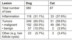
In dog toes, inflammatory processes with no indication of tumor growth were diagnosed in 118 cases (31.1%). This included various types of inflammation. Reactions to foreign material were frequently detected. These can cause extensive swelling of the toes. They can consist of introduced foreign materials like plant parts or hair that functions as endogenous foreign material following inflammatory processes or undirected growth. The type of inflammatory cells often indicates an additional bacterial infection. Some fungal infections as well as allergic reactions have also been observed.
If the inflammatory changes reach a chronic stage, increased granulation tissue is formed (Fig. 1).
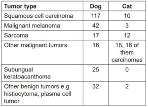
Tumorous lesions were diagnosed in 249 (65.5%) cases (Tab. 2).
In cat toes (n=87) inflammatory lesions were diagnosed in 33 cases (37.9%). Tumorous processes were detected in 51 submissions (58.6%) (Tab. 2).
Similar frequency distributions have been described in other studies of dogs and cats (Wobeser et al., 2007a and b).
Squamous cell carcinoma
Squamous cell carcinomas of the toes (Fig. 2+3) begin in the stratum spinosum of the epidermis of the claw or the sole. At first, they can appear clinically similar to inflammation of the nail bed. This can lead to lameness, swelling of the toe and ulcerations. The nail frequently appears softened and splintered. The tumor has a local invasive growth and, in advanced stages, leads to bone destruction.
In the literature, metastasis rates of 5 to 29% have been described.
Amputation of the toe is the therapy of choice. The risk of recrudescence is considered low if carried out correctly (resection in healthy tissue).
Standard and giant schnauzers have a breed predilection.
Subungual keratoacanthoma
Subungual keratoacanthomas, like squamous cell carcinomas, originate from the stratum spinosum of the epidermis. They form defined nodular growths with a central callus. Subungual keratoacanthomas can transform into malignant squamous cell carcinomas. They can displace bone tissue due to their expansive growth and can also cause lytic changes.
Melanoma
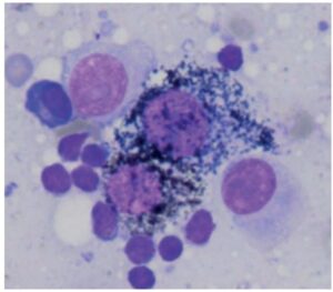
Melanocytic tumors of the toes (Fig. 4) can arise from melanocytes of the epidermis of the claws or the skin between the toes. Independent of their histological differentiation, they are often malignant on the toes. Despite amputation, metastasis is common, especially to the lymph nodes and the lung.
Carcinoma of the toes of the cat
In cats, tumors on the toe are frequently metastases of other primary tumors. Bronchial carcinomas in particular have a tendency to metastasize to the toes.
- Fig. 1: Mass on the dorsal aspect of the claw of a dog, which was histologically shown to consist of granulation tissue.
-
Fig. 2: Radiograph of the paw of a standard schnauzer with osteolysis of the distal phalanx of the 2nd toe. Histological examination showed a squamous cell carcinoma
Picture Credits: Dr. Sieberz, Kleintierklinik Ravensburg
- Tab. 1: Incidence of neoplastic and non-neoplastic lesions in submitted amputated toes from dogs and cats
- Tab. 2: Incidence of various tumors in amputated toes of submissions from dogs and cats
- Fig. 3: Infiltrative growth of a squamous cell carcinoma in a giant schnauzer (HE 10x).
- Fig. 4: Malignant melanoma of the toe of a dog (HE 400x)
Comments on pathological examinations
Cytological examination of masses of the toes are of very limited use, since they do not allow an evaluation of growth characteristics or the involvement of bone tissue. Many tumors on the toes are also superficially inflamed or ulcerated, so that cytology shows an inflammatory process and a tumorous process in deeper tissue layers is missed.
Inflammation can cause pleomorphism of the cells, which can resemble low malignancy neoplasias, making differentiation difficult.
Examination of biopsies before toe amputation may offer information on the type of tumor. If only inflammation is detected, this does not exclude the presence of a tumor in other areas of the toe.
Since bone tissue is always contained in amputated toes, these tissues must be decalcified in the laboratory prior to examination. This can take several days. In our laboratory, we first take soft tissues, if possible, and immediately embed them, allowing us to provide a preliminary result. This is particularly important since decalcification is associated with a loss of resolution of detail in the tissues. The resection margins of the bone tissue are evaluated after decalcification and are provided in a follow up report.
Conclusion for clinical practice
The amputation of swollen toes is a therapeutic option that can also be used to obtain a prognostically relevant diagnosis if the removed tissues are submitted for histological examination.
Literature
-
Heckel, Franziska (2012) Fallbericht: Subunguales Keratoakanthom bei einem Beagle – Es muss nicht immer ein Plattenepithelkarzinom sein. Kleintiermedizin 2/12, p. 82-88
-
Wobeser, B.K. et al. (2007a) Diagnosis and clinical outcomes associated with surgically amputated canine digits submitted to multiple veterinary diagnostic laboratories. Vet. Pathol. 44: 355-361
-
Wobeser, B.K. et al. (2007b) Diagnosis and clinical outcomes associated with surgically amputated feline digits submitted to multiple veterinary diagnostic laboratories. Vet. Pathol. 44: 362-365 (2007)
-
Kessler, M. (2013) Kleintieronkologie, 3. Auflage (2013): Diagnose und Therapie von Tumorerkrankungen. p. 428-431
