Leptospirosis is a zoonotic disease found worldwide. Leptospira is a gram-negative, helical shaped bacterium from the group spirochetes. Pathogen and non-pathogen species co-exist in the environment and are closely adapted to different vertebrate reservoir hosts which excrete the pathogen in the urine. Worldwide Leptospira are described in more than 150 mammal species. Dogs are predominantly infected with the species Leptospira interrogans. Pathogenic Leptospira are classified in different serovars due to different carbohydrate composition of the bacterial lipopolysaccharides, which in turn are grouped into serotypes. Although bivalent vaccines against the serovars L. icterohaemorraghiae and L. canicola have been available since the seventies, the number of leptospirosis cases has risen, so probably more serovars are involved in the infection.
Clinical Symptoms
The clinical manifestation of leptospirosis is quite complex and variable, and depends on both the pathogenicity of the serovar, the immune response of the host as well as the amount of bacteria taken. Leptospira are transmitted by direct contact (water, urine, contaminated soil, bites, placenta) and by oral ingestion of infected tissue (rodents). They multiply in the blood of the host on day 1 after infection and colonize the liver, kidney, lungs, spleen, CNS and the eyes. Symptoms vary according to the organ manifestation from severe to mild, unspecific symptoms. Most common symptoms are fever, anorexia apathy, icterus, hepatitis, coagulopathies, polydipsia/polyuria, vomitus, kidney failure, coughing and dyspnoea.
- Fig. 1
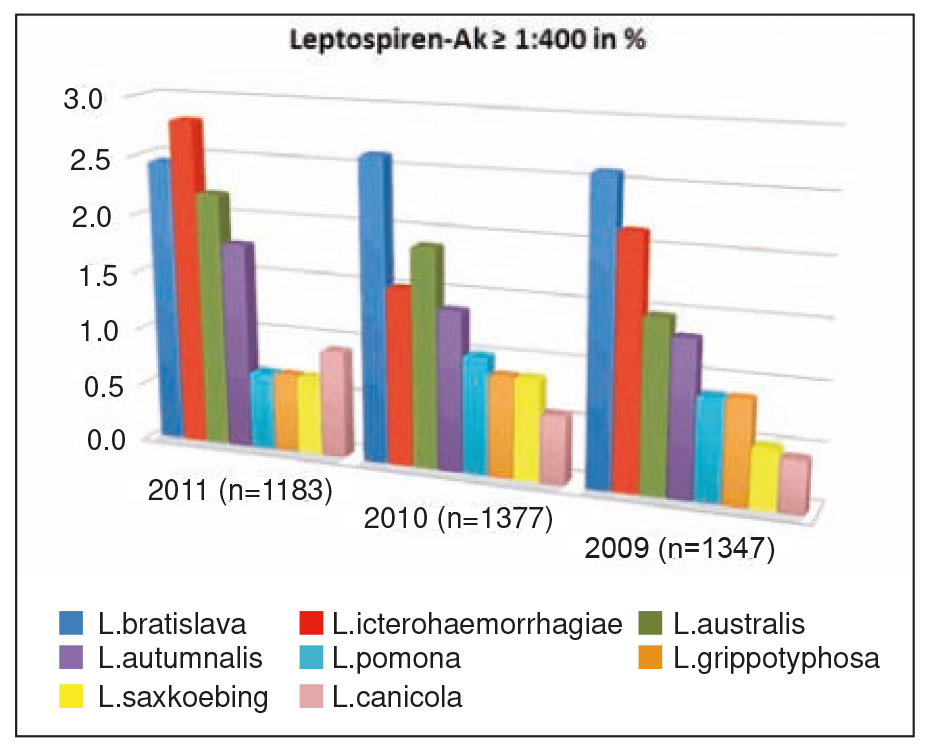
- Fig. 2: Leptospiren-Ak ≥1:400 in %
Diagnosis
The serological standard method for the indirect pathogen detections is the micro-agglutination test (MAT). Serial dilutions of the patient serum are incubated with living Leptospira serovars and then a possible agglutination is evaluated as an expression of the Ag/Ab-reaction microscopically.
A four-fold increase of antibodies (≥1:400) against a vaccine-serovar is considered diagnostic of an infection, as well as a positive titre against a nonvaccine serovar. Additionally, depending on the disease‘s organ manifestation, increases in liver enzymes, altered kidney values, leuco cytosis with a left shift, thrombocytopenia as well as DIC can be seen. Direct pathogen detection by a PCR test from urine may also be performed. This is only conclusive in positive cases and can be used to identify asymptomatic carriers. The urine PCR test can be false negative in patients who have received high doses of antibiotics.
Serovar prevalence in 3907 dogs
From 2009 to 2011 more than 35000 MAT-tests were performed at LABOKLIN. Vaccine status or medical histories were not known. The tested serovars were: L. icterohaemorraghiae, L. canicola, L. pomona, L. grippotyphosa, L. bratislava, L. autumnalis L. saxkoebing, L. australis und L. sejroe. Around 10% in 2009, 11% in 2010 and 12% in 2011 of all samples were positive (titres ≥ 1:400). Of the positive samples most had positive antibodies against L. bratislava, L. icterohaemorraghiae and L. australis, although a dynamic frequency was seen. In 2009 the most frequent serovars found were L. bratislava (~25%) > L. icterohaemorraghiae (~21%) > L. australis (~14%), which in 2010 changed to: L. Bratislava (~24%) > L. Australis (~18%) > L. icterohaemorraghiae (~14%) and in 2011 to L. icterohaemorraghiae (23%) > L. bratislava (~20%) > L. australis (~18%). The antibody prevalence of L. grippotyphosa varied from 2009 to 2010 only slightly (8.5% > 8.1%) but sank in 2011 to 5.5%. The percentage positive towards the serovar Saxkoebing was very consistent around 5%, with a transient increase in 2010 to 8%. Positive antibodies towards the (vaccine) serovar L. canicola rose from 5% (2009) to around 8% in 2011. None of the tested dogs had positive antibodies towards the serovar L. sejroe.
Further results
The highest antibody response (1:3200) was seen against L. bratislava, > L. autumnalis > L. australis > L. icterohaemorraghiae, although a dynamic change in frequency was observed. Whereas in the years 2009-2011 dogs positive towards only one serovar stayed the same (~4%), the number of dogs positive to two serovars slightly increased (1.3% in 2009 to 2.1% in 2011). Seropositivity to 3 and ≥ 4 serovars was also seen (0.3% and 0.6% respectively).
Summary
It is said that L. grippotyphosa and L. saxkoebing should be the most frequent serovars causing canine Leptospirosis in Southern Germany. In Eastern Germany it is said to be the L. pomona. Positive antibody titres towards both serovars could only rarely be determined from the samples examined by us. Also positive antibody titres (≥1:400) towards the vaccine serovar L. canicola were rarely seen, although they (vaccine serovars L. canicola and L. icterohaemorraghiae) were responsible for the largest percentage of detected low antibody titres (≤1:200). So when we take the high levels of detected antibodies as well as the frequency of the wild serovars L. Bratislava, L. Australis and L. Autumnalis into account, we can assume that a cross reaction to the vaccine serovars (L. Canicola, L. Icterohaemorraghiae/L. Copenhageni) is possible, but unlikely. It is therefore now assumed that further serovars are involved in the infections, which earlier were considered as „rare“. For the clinician, Leptospirosis should still be considered as an important differential diagnosis, even in vaccinated patients, if suspicious clinical symptoms occur.


