Pet reptiles are frequently presented to the practice for various types of skin lesions. There are many possible causes for these lesions and they are often multifactorial. These include, first and foremost, husbandry issues, such as inappropriate temperature or humidity, unsuitable substrate or furnishings or poor hygiene. A detailed clinical history and an exact diagnosis are essential for successful treatment.
The purpose of this Laboklin aktuell is to give you a brief overview of the most important infectious causes of skin lesions (excl. parasites). Mixed infections are common and many factors influence how infective dermatitis progresses. A difinitive diagnosis often requires tests such as cytology and histopathology combined with direct pathogen detection and interpretation of results in the context of the clinical presentation.
Viral causes
There are a few viruses which are frequently found in association with skin lesions in reptiles. In some cases, they are considered the primary cause, while in others, their role in the development and progression of the disease is less clear.
Iridoviridae
Iridoviruses of the genera Ranavirus and Iridovirus have been found in reptiles. Both have been described in association with of skin lesions, especially in lizards.
-
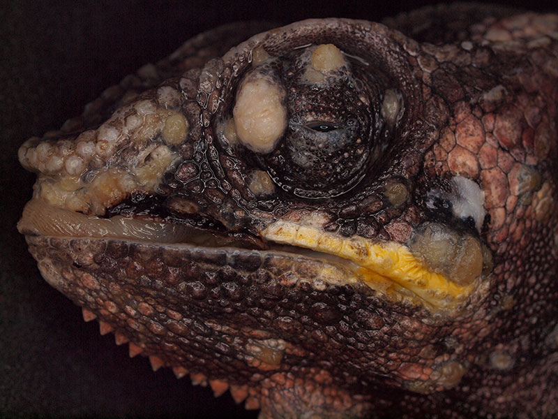
Fig. 1: Panther chameleon (Furcifer pardalis), multiple papillomas.
Photo credits: Dr. Janosch Dietz, Laboklin GmbH
-
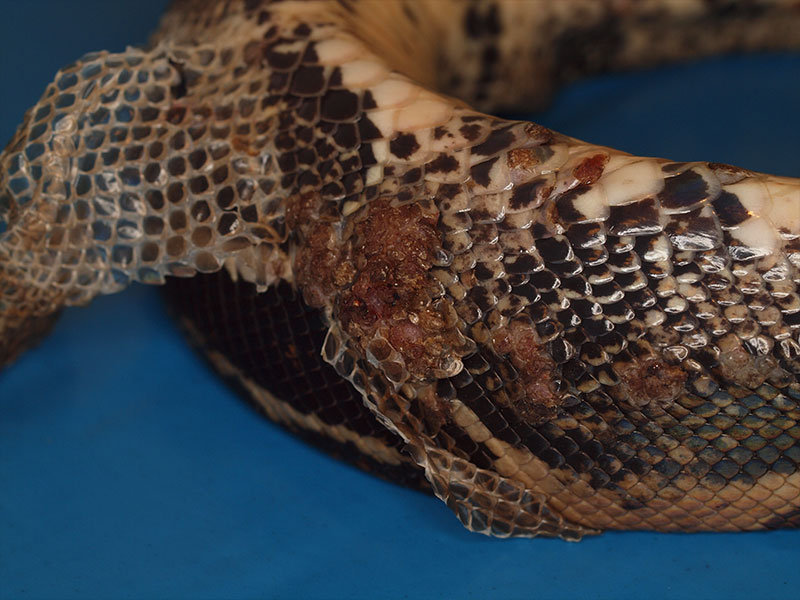
Fig. 2: Boa constrictor, papillomas
Photo credits: Dr. Janosch Dietz, Laboklin GmbH
Ranaviruses
Ranaviruses can infect amphibians, fish and various reptile species. In reptiles, they have most commonly been described in chelonians, but skin lesions caused by ranaviruses are mainly found in lizards. They can manifest as subcutaneous neck oedema, dermatitis or abscesses. Severe to fatal courses, depending on various factors, as well as inapparent infections have been described.
Iridoviruses (Invertebrate Iridovirus – IIV)
IIV are primarily detected in lizards. They regularly occur in feeder animals (e.g. crickets) and it is suspected that they are transmitted from insects to reptiles. It is unknown whether IIV has an impact on the health of infected reptiles. Nevertheless, they are regularly detected there, for instance in skin samples, especially from lizards, with various skin lesions such as pox-like lesions or loss of scales.
Herpesviridae
Herpesviruses are mainly found in chelonians. Various viruses with different host specificities play a role there. Some of them are associated with skin lesions. Herpesvirus infections in sea turtles often present with skin lesions, such as fibropapillomatosis. Papillomatous lesions have also been described in aquatic turtles and lizards with herpesvirus infections. Individual cases of ulcerative skin lesions have been described in reptiles. In crocodiles, herpesviruses are associated with lymphocytic infiltration of the skin.
Papillomaviridae
Papillomaviruses are very host-specific and tissue-associated. Infections cause the formation of papillomas and skin growths. They have been described in individual cases in different reptile species.
Poxviridae
Poxviruses are mostly described in crocodiles. They have also been detected in various chelonian species suffering from skin lesions. In one lizard, ulcerative skin lesions were reported.
Reoviridae
In snakes and lizards, reoviruses have mainly been detected in diseases of the intestinal tract, the central nervous system and the lower respiratory tract. Reovirus infections have also been described in papillomas as well as in necrotising and ulcerative dermatitis.
Arenaviridae and inclusion body disease (IBD)
Reptilian arenaviruses are assigned to the genus Reptarenavirus. So far, species of this genus have only been found in snakes. In boas and pythons, they cause inclusion body disease (IBD). IBD can present in very different ways, particularly through neurological, gastrointestinal and respiratory signs. Skin lesions are also regularly observed, especially in boas. They can vary in severity and range from moulting disorders to extensive dermatitis.
Mycological causes
Dermatomycoses are often caused secondarily and only appear after immunosuppression.
Clinically, they may resemble a bacterial infection, in which case bacterial and fungal culture is an important tool for differentiation. However, there are some primary pathogenic fungi that are regularly detected in reptiles, especially from the order Onygenales.
Clinical infection is generally characterised by localised, crusty, yellow to brown skin lesions, blistering and hyperkeratotic to necrotising skin areas. In snakes, infections with Ophidiomyces ophidiicola play a role in wild animals as well as in domestic and zoo animals. This fungus causes “snake fungal disease” or ophidiomycosis and seems to have a broad host range in snakes. Infected animals commonly develop crusty dermatitis, which is often distributed on the head. Severe mycoses are also possible. In some animals, changes are milder, e.g. colour changes or the formation of subcutaneous nodules. Less severely affected animals may be temporarily free of signs after moulting. In lizards, especially agamas, infections with fungi of the genus Nannizziopsis, particularly species like N. guarroi, N. dermatitidis or N. vriesii, cause severe skin changes and systemic disease. They were formerly called CANV (Chrysosporium anamorph of Nannizziopsis vriesii) and the associated disease was referred to as “yellow fungus disease”. However, the term nannizziomycosis is preferred. Clinically, crusty dermatitis is seen, which can also invade deeper tissue. There have also been individual reports of infections with related dermatophytes in connection with dermatitis in crocodilians and turtles.
Bacteriological causes
Bacterial skin infections in reptiles are usually the result of systemic diseases or poor husbandry. Clinically, they may present as dermatitis or abscesses. Bacterial pathogens can colonise the skin primarily or by haematogenous spread. The spectrum of pathogens found in skin lesions in reptiles can vary significantly. The clinical relevance of bacteria detected in skin lesions must be assessed on a case-by-case basis. Only a few primary pathogenic bacteria are known to cause dermatitis. Devriesea agamarum, a gram-positive bacillus, has been isolated in lizards, especially in spiny-tailed lizards. This bacterium is associated with dermatitis, mostly chronic proliferative and scaly changes, and septicaemia. There are also various disease complexes involving different bacterial species in which typical skin changes occur. In turtles, “septicaemic cutaneous ulcerative disease” (SCUD) occurs which affects the shell. This syndrome is often seen with a mixed infection of various potentially pathogenic bacteria, especially Citrobacter spp.
Lisa Schüler and PD Dr. Rachel Marschang
Table: Common causes of infectious dermatoses in pet chelonians, lizards and snakes
| Pathogens | Hosts | Clinical signs concerning the skin | Diagnosis (methods and material) |
| Viruses | |||
| ranaviruses | chelonians | occasional involvement of the skin: subcutaneous oedema, dermatitis or abscesses; redness with petechiae; skin ulcerations | PCR*: swab without medium, biopsy (fresh or with a small amount of NaCl), skin scraping, tissue (esp. liver) histopathology*: biopsy or tissue (esp. liver) in formalin |
| lizards | skin lesions (multiple, grayish-brownish, crusty to partly ulcerative) | ||
| invertebrate iridoviruses (IIV) | lizards | pox-like skin lesion, loss of scales | |
| herpesviruses: different strains e.g. Terrapene herpesvirus 2 |
chelonians | ulcerative lesions on skin and shell, papillomatous proliferative skin changes |
PCR*: biopsy (fresh or with a small amount of NaCl) and/or skinscraping (esp. papillomas), swab without medium (lesions), tissue histopathology*: biopsy in formalin |
| lizards | papillomas | ||
| papillomaviruses | chelonians, snakes and lizards | proliferative skin lesions, papillomas, squamous epithelial carcinoma | PCR*: biopsy (fresh or with a small amount of NaCl) and/or skinscraping (esp. papillomas or skin growths), swab without medium (lesions) histopathology*: biopsy in formalin |
| poxviruses | chelonians and lizards | papular skin changes, vesicles on skin and shell, oedema in neck area | PCR: biopsy (fresh or with a small amount of NaCl) and/or skin scraping, swab without medium (lesions), tissue histopathology*: biopsy in formalin |
| reoviruses | lizards | papillomas, necrotising and ulcerative dermatitis | PCR*: biopsy (fresh or with a small amount of NaCl) and/or skin scraping (esp. papillomas), swab without medium (lesions), tissue |
| reptarenaviruses | pythons and boas | inclusion body disease (IBD); skin lesions | PCR*: biopsy (fresh or with a small amount of NaCl), skin scraping, tissue histopathology*: biopsy in formalin, blood smear (esp. boas), tissue (esp. brain, pancreas and liver) |
| Bacteria | |||
| Devriesea agamarum | lizards | mostly chronic proliferative, exudative and crusty dermatitis (partly scaly), cheilitis | histopathology*: biopsy in formalin
cytology*: impression smear of the skin lesion or skin scraping aerobic and anaerobic culture test* |
| Austwickia chelonae, A. chelonae-like and (formerly) Dermatophilus-like | tortoises and aquatic turtles, lizards and snakes | skin lesions; dissemination into deeper tissue and subsequent granuloma formation possible | |
| other species (aerobic and anaerobic) | all species | dermatitis, abscesses | |
| Fungi | |||
| Ophidiomyces ophidiicola | snakes | “snake fungal disease” or ophidiomycosis: crusty dermatitis | PCR (Ophidiomyces ophidiicola*): swab without medium, biopsy (fresh or with a small amount of NaCl), skin scraping
histopathology*: biopsy or tissue in formalin cytology*: impression smear of the skin lesion or skin scraping culture test*: swab with medium; alternatively also biopsy (with a small amount NaCl) or skin scraping |
| Nannizziopsis spp. | lizards | “yellow fungus disease”: crusty dermatitis | |
| Paranannizziopsis spp. | snakes and lizards | crusty dermatitis | |
| Emydomyces testavorans | chelonians | ulcerative shell lesions, partly severe | |
| other facultative pathogenic fungi | various species | dermatitis | |
*a service provided by Laboklin


