In Europe, cowpox virus is the most relevant representative of the genus Orthopoxvirus in the family Poxviridae. Cowpox viruses have zoonotic potential and occur endemically in Europe as well as in Northern and Central Asia.
Numerous species have been identified as hosts, including humans, pet rats and cats. Moreover, there have also been individual cases in dogs, primates, elephants, rhinos and exotic felids. Wild rodents, especially voles (e.g. bank voles), are considered the natural reservoir of cowpox virus. Domestic animals with close contact to humans pose the greatest zoonotic risk.
Feline cowpox virus infections occur rather rarely. However, they are often not recognised clinically and have become an important route of transmission from animals to humans in recent years.
Infections are typically observed in outdoor cats. They mainly occur in late summer and autumn, since this is the time the rodent population is at its peak.
The primary infection is usually the result of local bite wounds from infected prey, especially on the forelimbs, the chest as well as in the face. Initially, these are small local changes which can worsen due to secondary infections. Multiple/ generalised skin lesions may develop within a few weeks as a result of leukocyte-associated viraemia. Systemic changes are uncommon in immunocompetent cats, but fatal pneumonia is seen in kittens, immunocompromised animals and exotic felids (cheetahs).
It must be pointed out that there have been individual case reports which describe respiratory signs alone or only secondary, atypical skin lesions.
In these cases, diagnosis could only be made after taking a biopsy of the affected site or through further examination.
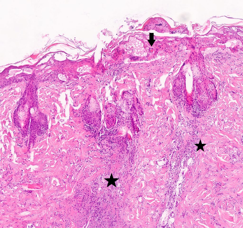
- Fig. 1: Skin biopsy with severe dermal necrosis (star), epidermal hyperplasia with oedema formation and intracorneal intracytoplasmic inclusions. 4x HE.
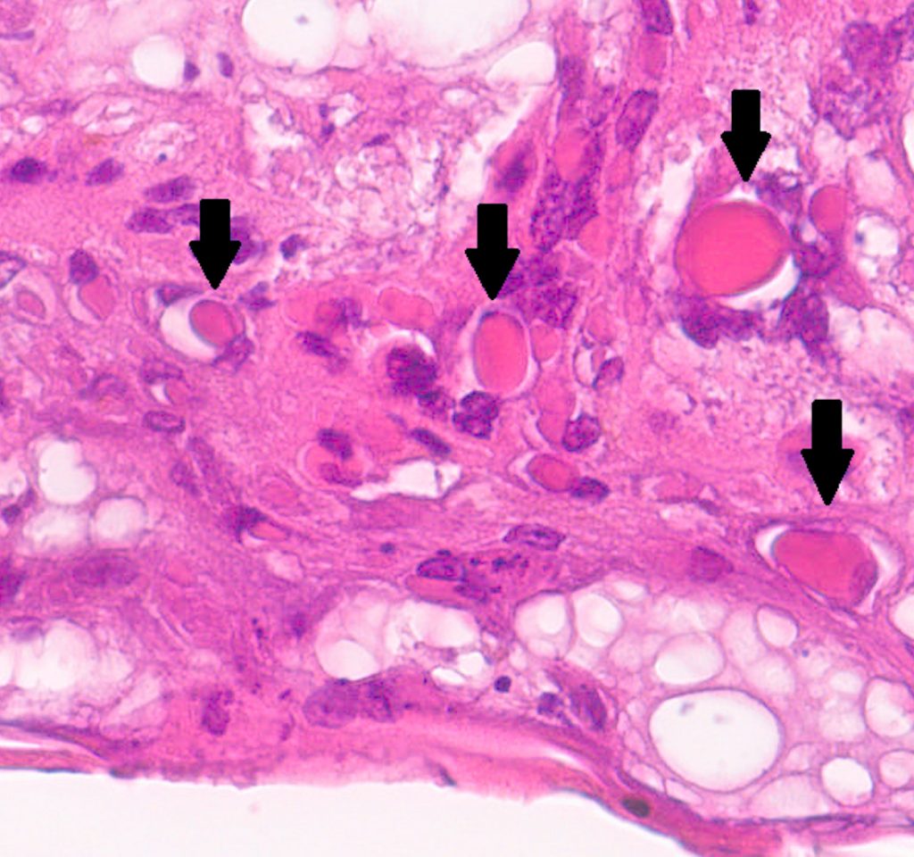
- Fig. 2: Skin biopsy. Detection of keratinocytes with intranuclear eosinophilic inclusion bodies (arrows). 40x HE.
Clinical picture
Clinically, most cases are reported to be outdoor animals which have poorly healing or proliferative skin lesions, especially on the head and chest or on the forepaws and ears. These lesions first present as well circumscribed, initially small hyperaemic patches or macules which grow to become papules and nodules. As a characteristic feature, they develop a central ulceration with depressed necrosis. These kinds of changes may also occur on the tongue and the oral mucosa. Generally, the crusty lesions heal with scarring within 3 – 12 weeks. Relapses do not usually occur, but healing may be delayed if there is a secondary infection (bacterial or mycotic). Systemic changes may be observed during the viraemic phase, but present as mild. However, if feline cowpox virus occurs as a co-infection in immunocompromised animals, during an infection with feline immunodeficiency virus (FIV), feline leukosis virus (FeLV) or feline parvovirus, fatal complications may arise. Fatal pneumonia can also be observed due to iatrogenic immunosuppression (corticosteroid therapy).
It should be noted that unusual manifestations of cowpox virus infections can occur as well; these have only recently been described in the literature (Jungwirth et al., 2018).
These patients presented because of other symptoms (e.g. trauma) and subsequently developed skin lesions characterised by local oedema formation and hyperaemia of the skin as well as mild plaque in the limbs.
Another unusual case was a young cat (Schöniger et al., 2007). This cat was presented with purely respiratory signs with acute onset of dyspnoea followed by pneumothorax. A biopsy was taken in this case and showed necrotising to proliferative bronchointerstitial pneumonia with pneumocytes, which had indicative intracytoplasmic inclusions. The diagnosis of „feline cowpox virus“ was made by molecular biological investigation.
Detection of pathogens
The methods of choice for pathogen detection are tissue biopsy with subsequent pathohistological examination and molecular biological examination using polymerase chain reaction (PCR) for the detection of viral DNA.
It should be ensured that the biopsy is taken from the edge with epidermal and dermal parts, as characteristic structures (inclusion bodies) can be detected in them. Diagnosis is often easier in early lesions, as extensive necrosis with tissue loss may dominate in late lesions.
Histological examination typically reveals severe epidermal and adnexal necrosis (Fig. 1) with large, intracytoplasmic, eosinophilic inclusion bodies (Fig. 2).
Histological examination is recommended to confirm a suspected feline cowpox virus infection and to clarify a secondary process or other possible differential diagnoses.
Differential diagnoses
Non-healing or poorly healing proliferative crusty lesions on the head, the ears as well as the limbs can have numerous infectious and non-infectious causes in cats and must be differentiated from feline cowpox virus infection.
Differential diagnoses may include bacterial and mycotic infections, but also an autoimmune process such as pemphigus foliaceus, which presents histologically with subcorneal and intracorneal neutrophilic pustules with acantholytic keratinocytes (rounded and hypereosinophilic keratinocytes). These dead acantholytic cells may be misinterpreted as eosinophilic inclusion bodies.
Eosinophilic granuloma complex may be another differential diagnosis. Histologically, within severe cases a marked dermal eosinophilia with typical „flame figures“ is observable. However, neoplastic processes (e.g. Bowenoid in situ carcinoma or squamous cell carcinoma) are also possible differential diagnoses. Furthermore, other viral infections, such as feline herpesvirus 1, should be considered as a potential cause as well. Feline herpesvirus 1 may histologically present very similar to feline cowpox virus, except for the fact that it has characteristic intranuclear basophilic inclusion bodies, whereas feline cowpox virus shows intracytoplasmic eosinophilic inclusions. However, inclusion bodies can often not be detected histologically or are very difficult to identify.
Treatment and management
No specific therapy is available to treat cutane cowpox virus infections.
Various supportive measures, such as thorough cleaning and, if required, antibiotic treatment, may be indicated to prevent secondary bacterial infection.
Patients with a severe course of the disease need intensive supportive therapy. Treatment with glucocorticoids is contraindicated.
Isolating the cat until the lesions are completely healed and appropriate hygiene measures are advisable. The virucidal disinfectants recommended by the German Veterinary Medical Society (alcohol and ethyl ether are not suitable) have proven to be effective. Additionally, inactivation at >80 °C is possible.
It should also be kept in mind that virus particles in crust material as well as in dry swabs can remain active for a longer period of time (months) at room temperature.
In general, contact with children and immunocompromised persons should be avoided until the lesion is completely healed – especially because of the high viral load in the crust material and in the secretions of skin wounds of infected animals.
Conclusion
Feline cowpox virus infection is a rare but sporadic zoonosis that particularly affects outdoor cats. If clinically there are poorly healing, raised papular to pustular skin lesions with central depression, especially on the head and chest and on the forelimbs, cowpox virus infection should be considered.
Moreover, it is recommended to inform owners about the zoonotic potential, especially in the case of immunocompromised contacts, and further work-up should be done (histological examination and/or molecular biological examination).
Please note that according to the legislation on epizootic diseases, Orthopoxvirus infections are notifiable and must therefore be reported to the competent veterinary authority.
Dr. Nicole Jungwirth


