The diagnosis of thyroid hormone disorders plays a significant role in small animal medicine, especiall in dogs, cats, and guinea pigs. However, birds and reptiles can also exhibit thyroid disorders, often due to causes like thyroiditis or iodine deficiency.
Hypothyroidism has been reported in various bird species, including grey parrots (Psittacus erithacus), amazons, macaws, parakeets, and others.
For instance, a case involving a hyacinth macaw (Anodorhynchus hyacinthinus) revealed ulcerative dermatitis and valvular endocarditis (Huynh et al., 2014). Additionally, feather loss, obesity, hypercholesterolemia, and non-regenerative anemia have been documented in affected birds (Oglesbee, 1992). On the other hand, hyperthyroidism, often resulting from hormone-producing tumors, is rare in birds.
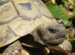
Tortoises with hypothyroidism exhibit anorexia, lethargy, and myxoedema of the skin around the head, neck, and forelimbs. Due to the thyroid gland’s location at the base of the heart, swelling in the neck area is rarely observed. Giant tortoises, such as the Galapagos tortoise (Chelonoides nigra) and Aldabra tortoise (Aldabrachelys gigantea), are particularly affected. The underlying cause is usually an iodine deficiency in the diet or the consumption of goitrogenic plants, such as pak choi, broccoli, cabbage, and soya. A case of hyperthyroidism has also been documented in a green iguana (Iguana iguana), triggered by a thyroid adenoma. This condition led to weight loss, polyphagia, hyperactivity, increased aggression, loss of dorsal spines, tachycardia, and swelling in the neck area (Hernandez-Divers et al., 2001).
The diagnosis of hypothyroidism in birds and many reptile species can be particularly challenging, as thyroxine levels (tT4) in the blood are typically very low within the normal range. As a result, they often fall below the detection limits of commonly used analyzers (e.g. < 0.12 µg/dL). In birds, TSH stimulation tests using bovine and human TSH have also shown success. However, routine TSH measurement is not currently possible due to the species-specific molecular structure, and free T4, T3, and fT3 are rarely determined diagnostically.
Laboklin has developed an LC-MS method that reliably measures low tT4 levels in birds and reptiles, with a detection limit of < 0.02 µg/dL. This method has been tested and validated using blood samples from various species of turtles, lizards, snakes, and psittacids and is now available for routine diagnostics under service number 1089. However, due to the large species variability, no reference values can be provided.
-
-
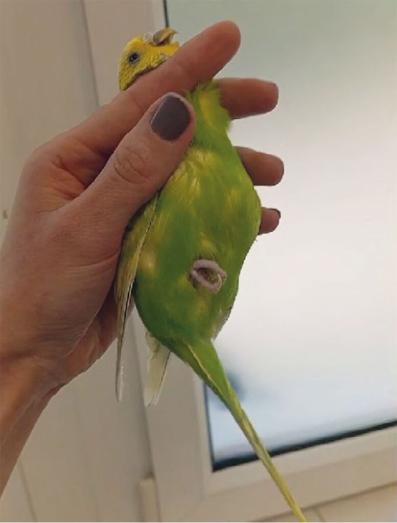
Fig. 1: Budgerigar (Melopsittacus undulatus) undergoing treatment for hypothyroidism
Image source: Dr. Christoph Leineweber

-
-
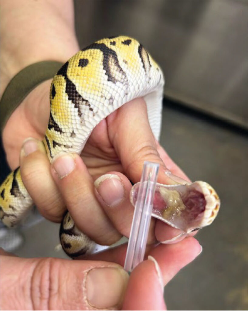
Fig. 2: Royal python (Python regius) exhibiting respiratory symptoms and positive mycoplasma detection
Image source: Dr. Christoph Leineweber

Mycoplasma Infections in Snakes
In turtles, mycoplasma infections associated with upper respiratory tract disease (URTD) and symptoms such as nasal and ocular discharge, conjunctivitis, and eyelid oedema have been well-documented and are widespread. In alligators and crocodiles, mycoplasmas can lead to septicaemia and arthritis. But what role do mycoplasmas play in snakes?
Until now, there have only been isolated reports of mycoplasma infections in snakes. A recent study from 2021 by colleagues at Laboklin revealed that mycoplasmas are relatively rare in pythons, but they occur more frequently (n = 271, 60% positive) (Racz et al., 2021). Previous case reports have described symptoms such as tracheitis, pneumonia, stomatitis, and anorexia associated with positive mycoplasma detection in pythons (Penner et al., 1997; Schmidt et al., 2013; Marschang et al., 2016; Magalhães et al., 2021). However, the exact link between mycoplasmas and clinical disease in snakes remains unclear.
Interestingly, the mycoplasmas described so far have shown a close relationship to Mycoplasma [Mycoplasmopsis] agassizii, which is known to cause severe diseases in turtles (Penner et al., 1997; Marschang et al., 2016; Magalhães et al., 2021; Racz et al., 2021). There has been little research on the occurrence of mycoplasmas in snakes from other families. A 2021 study described the detection of mycoplasmas in four boas (Boa constrictor) and one viper (Bothrops atrox) for the first time (Magalhães et al., 2021).
For the diagnosis of a mycoplasma infection, suitable sample materials include throat swabs (without medium) or a nasal rinse sample.
You are also welcome to send tissue samples.
For pythons, we offer a respiratory/neurological PCR profile (service number 8262), which includes the detection of mycoplasmas. The mycoplasma PCR can also be requested as an individual service (service number 8088); it is also suitable for various other snake species.
Dr. Christoph Leineweber