Alopecia in dogs can have many causes. There may be alopecia secondary to scratching, licking or pulling of hair in dogs with pruritus. There are also numerous non-inflammatory causes of hair loss:
- Congenital or acquired genetic defects: congenital alopecia, hypotrichosis, follicular dysplasias (e.g., colour dilution alopecia), alopecia X and pattern baldness
- Interruption of the hair cycle: as seen in endocrinopathies, anagen and telogen effluvium and post-clipping alopecia
- Clinically no recognisable inflammation, but nevertheless of inflammatory origin: alopecia areata, ischaemic dermatopathy/vasculitis, scarring alopecia, traction alopecia
One of these diseases, colour dilution alopecia (CDA), also known as blue dog or blue Doberman syndrome, is becoming more and more relevant.
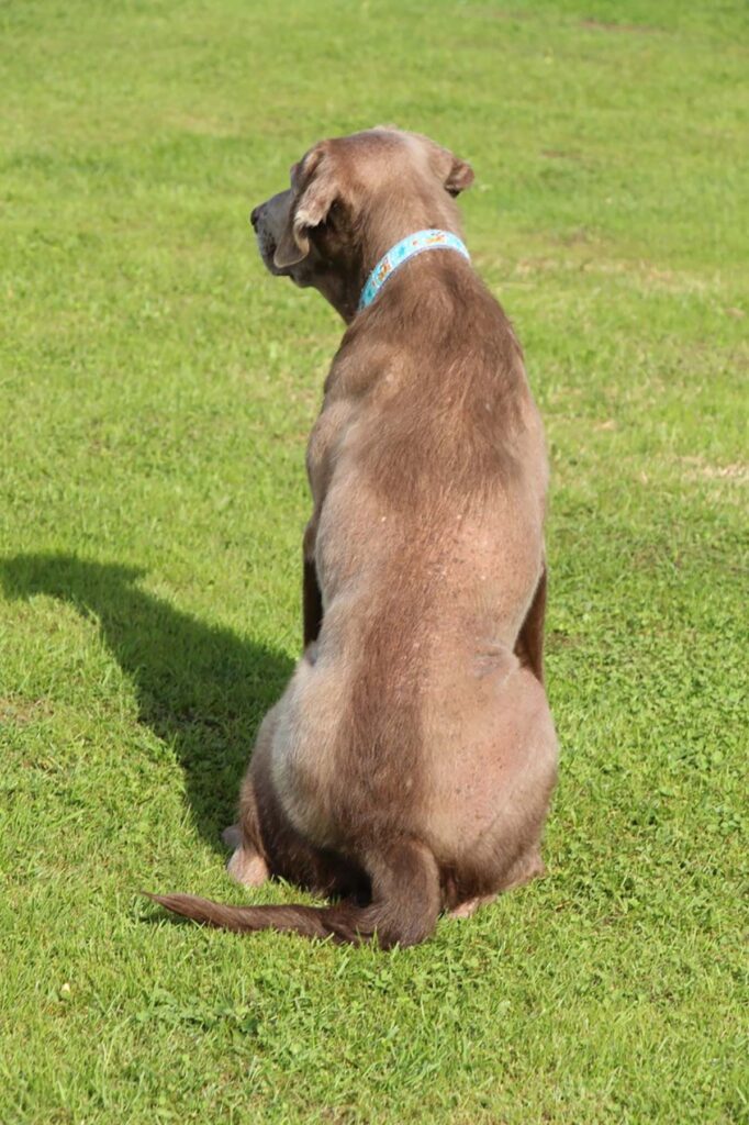
Dog breeders have noticed that specific coat colours are very popular with dog owners, even though they are considered undesirable according to the Fédération Cynologique Internationale (FCI) standard for the breed in question. The FCI-associated kennel clubs generally classify these colours as off-colours, which is why they are bred outside these associations. Labrador Retrievers (charcoal and silver, Figure 1), French Bulldogs (blue), as well as Chihuahuas, Prager Rattlers and the American Staffordshire Terriers (Figure 2) are increasingly affected. Colour dilution is recognised according to the FCI breed standard for the last three. Most puppy buyers are unaware that precisely these coat colours can often lead the dog to the vet in its lifetime.
Blue (charcoal) and lilac (silver) are based on a dilution of colour often associated with colour dilution alopecia. This condition causes progressive alopecia due to an alteration in the formation and storage of pigments in the hair. It is caused by a genetic variant of the dilute gene. Dilute means «attenuated» and refers to the lightening of black pigment to blue or brown pigment to lilac. The altered refraction of light results in an attenuated (diluted) colour tone (black becomes blue, brown becomes lilac).
Classically, CDA is particularly well known in the blue Doberman.
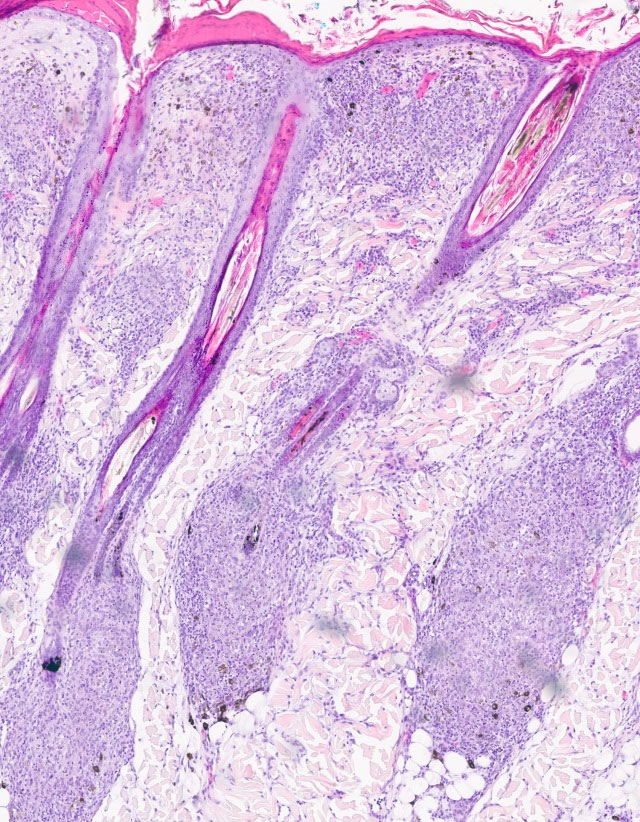
Clinical signs: The first clinical signs of CDA may appear as sparsely furred ears and tails, overall coat can also appear thinner than usual for the breed. At a few months of age, CDA may manifest as progressive alopecia of the trunk. Reduced hair density or hypotrichosis is most commonly seen in puppies (3 – 12 months old) of predisposed breeds. However, there are cases where alopecia occurs later in life. Alopecia progresses with age. Almost pathognomonic is the alopecia of areas of dilute colour adjacent to hairy areas of white or pheomelanin (yellowish pigment) pigmented hair. Dogs with CDA are prone to secondary bacterial infections (folliculitis and bacterial furunculosis, Figure 6). These dogs often suffer from recurrent cutaneous infections throughout their lives.
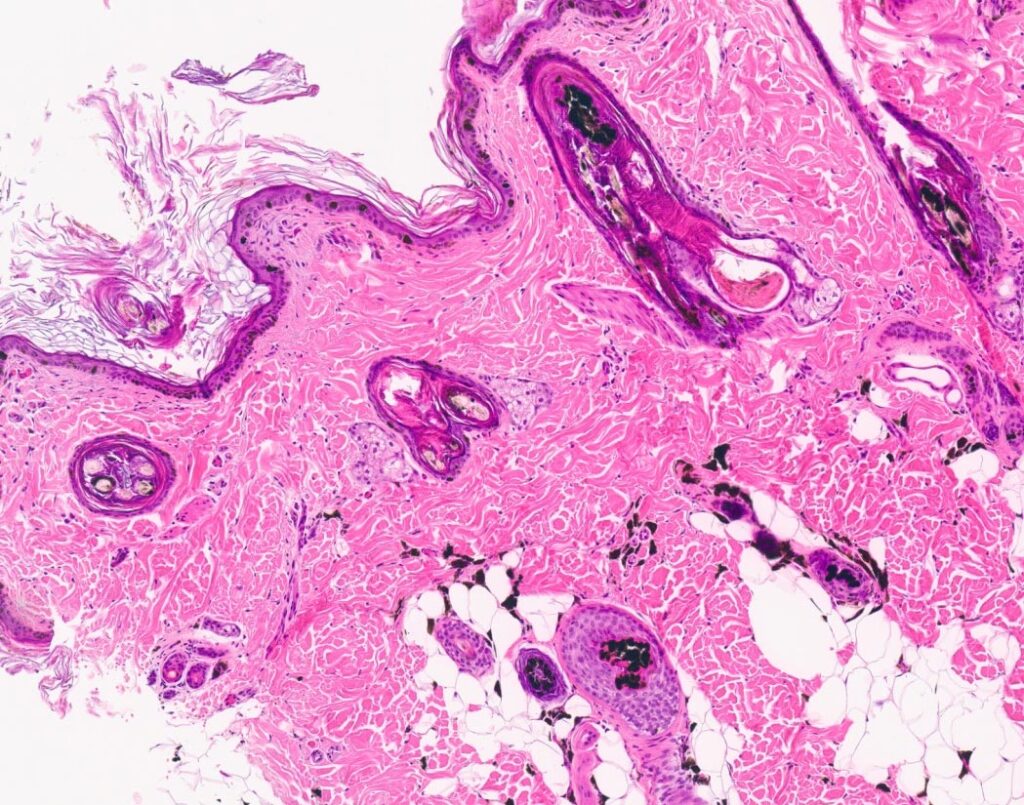
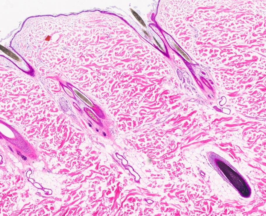
Pathogenesis: CDA is associated with colour attenuation due to a recessive genetic variant of the MLPH gene. This variant in homozygous genotype (d/d) leads to a defect in the transport of melanosomes from the centre of the cell to the periphery, resulting in clumping of melanin granules (Figures 4 and 5). The result is a defective pillar structure, which causes the hair to break easily.
Diagnosis: The diagnosis of CDA is based on a combination of the clinical picture (alopecia), microscopic examination of the hair and histopathology. Trichography shows the presence of giant melanosomes (melanin clusters) in the hair shaft’s cortex, with defects and fractures of the cuticle. Histopathological findings include hyperkeratosis and infundibular keratosis with possible melanin clusters in the epidermis; pigment incontinence with the presence of perifollicular and peribulbar melanophages; dysplastic hair follicles and shafts with irregular pigmentation and melanin deposits at all levels of the hair follicle, including the bulb (Figure 4).
Treatment: CDA cannot be cured, but the condition can be improved by controlling secondary infections. Fatty acid supplementation may improve the coat but does not influence hair growth. In some cases, melatonin may stimulate hair growth, but usually only for the duration of the treatment. Potential risks should be assessed before administration, especially if the animal has not reached sexual maturity, in pregnant and lactating bitches and in patients with liver and kidney disease. Dogs with CDA should not be used for breeding.
Genetic tests: There appears to be a breed-specific risk of CDA, as not all dilute colour dogs develop alopecia. The classically bred German Weimaraner is exclusively dilute brown (lilac), and CDA does not appear to affect these breeding lines. The same is true for the blue colour of the Great Dane. In the Doberman, however, the proportion of dilute colours with alopecia was so high that the blue colour was removed from the FCI breed standard.
Genetic testing cannot predict whether a dilute colour dog will develop CDA. Genetic tests exist for the three testable variants of the d allele (d1, d2 and d3). Still, no genetic tests are yet to distinguish between «dilute colour without CDA» and «dilute colour with CDA». Other variants are suspected but not yet identified. The wild form of D(N) is dominant, meaning all dogs with a dominant D(N) allele will phenotypically have a non-dilute coat colour. However, mating two dogs with the D/d (N/d) genotype may result in offspring with the d/d genotype. These will have a diluted coat colour because they do not produce regular pigment granules but smaller, clumped ones. For breeders of breeds affected by CDA, it is essential to use genetic testing to identify carriers of the d allele in the parents. Currently, CDA can only be diagnosed histologically by skin biopsy.
Dr Barbara Gruber, Dr Anna Laukner
-
Fig. 1: Silver Labrador Retriever
Source: envatoelements
-
Fig. 2: Blue Staffordshire Terrier
Source: Shutterstock
-
Fig. 3: Patient with CDA, Silver Labrador Retriever
Source: Dr Anna Laukner
-
Fig. 4: Histology CDA, HE staining, hair follicle with intra and extrafollicular melanin clumps. Dachshund, 3 years old
Source: Laboklin
-
Fig. 5: Histology, HE staining, normally pigmented black hair. Bernese Mountain Dog
Source: Laboklin
-
Fig. 6: Histology CDA with folliculitis and furunculosis. HE staining. Blue Weimaraner, 2 years old
Source: Laboklin
Diagnostic possibilities for the detection of dilution:
- D-locus d1
- Rare D-locus variants (d2, d3)
Diagnostic options for the detection of CDA
- Histopathological examination of a skin biopsy


