The acute-phase response (APR) is an early systemic reaction to inflammatory stimuli, infections, and tissue damage. The synthesis of specific acute-phase proteins (APPs) in the liver is regulated by proinflammatory cytokines (IL-6, IL-1β, TNF-α), with their concentrations in the blood rising or falling during an APR. APPs are valuable markers for detecting and monitoring the progression of inflammatory processes as well as neoplastic diseases. The APR occurs significantly earlier and more specifically than changes in leukocyte counts.
APPs are classified based on the magnitude of their response into major (10–100-fold increase), moderate (2–10-fold increase), and minor (<2-fold increase).
Measurement of major APPs is particularly useful for early diagnosis and monitoring of diseases associated with an APR. In dogs, C-reactive protein (CRP) is the primary major APP, whereas in cats, serum amyloid A (SAA) predominates. Haptoglobin (Hp) and α1-acid glycoprotein (AGP) are considered moderate to minor responding APPs. Albumin, transferrin, and paraoxonase are negative APPs, whose concentrations typically decrease during an acute-phase response. The behaviour of the respective APPs during the APR is summarised in Table 1.
-
-
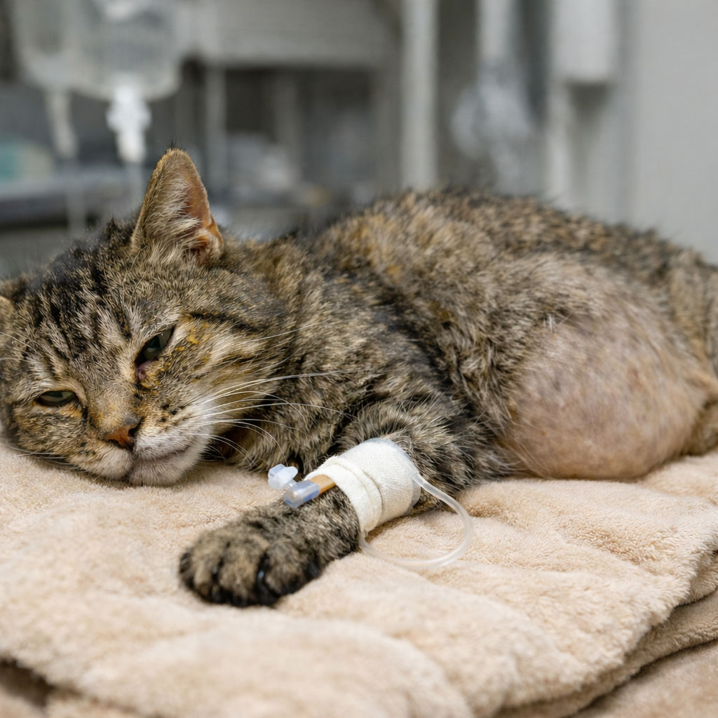
Fig. 1: Cat with FIP
Picture credits: Laboklin
Dogs – CRP as the Gold Standard
As a major APP, C-reactive protein (CRP) increases within 4–24 hours following the triggering insult, rising up to 50–100-fold, reaches its peak after 1–2 days, and declines rapidly with effective therapy.
Due to this dynamic behaviour, CRP is particularly well suited for early detection, monitoring disease progression, and assessing therapeutic success.
Elevated CRP levels are observed in a wide range of inflammatory and immune-mediated processes, including bacterial infections, parasitic diseases, autoimmune disorders, neoplasms, and post-traumatic or postoperative changes. In dogs with acute Babesia canis infection, CRP shows a strong correlation with clinical severity and haematological parameters.
Within the framework of antimicrobial stewardship, it has been shown that antibiotics can be discontinued once clinical improvement is observed and CRP concentrations have returned to normal, which significantly shortens the treatment duration for many diseases.
In systemic mycoses, such as pulmonary coccidioidomycosis, CRP in combination with haptoglobin (Hp) also demonstrated predictive value for remission. However, CRP can be elevated in the absence of inflammation, for example during extreme physical exertion or pregnancy, so its interpretation must always take the clinical context into account.
Acute-Phase Index (API) – a Combined Marker
Recent research has combined positive APPs (CRP, Hp) and negative APPs (albumin, optionally PON-1) into an acute-phase index (API). This index reflects the overall activity of inflammation. Dogs with malignant tumours and a high API had a significantly poorer prognosis.
Table 1: Overview of changes in acute-phase proteins and leukocyte counts over time following an inflammatory stimulus in dogs and cats.
| Time after Inflammatory Stimulus | Dog – e.g., CRP, SAA |
Dog – Leukocyte Count |
Cat – e.g., CRP, SAA |
Dog – Leukocyte Count |
| 0–6 h | slight increase at the start (hepatic synthesis begins within a few hours) | usually still within the reference range | slight increase at the start | usually still within the reference range |
| 6–12 h | marked increase measurable | first tendency to rise possible, often still borderline | marked increase measurable | first tendency to rise possible, often still unremarkable |
| 12–24 h | strong increase, values usually clearly pathological | leukocytosis or leukopenia often visible | strong increase, clearly pathological | more frequent leukocytosis, sometimes stress leukogram |
| 24–48 h | peak of APR – highest concentrations | further increase or plateau of leukocytes | peak of APR | further increase or plateau of leukocytes |
| 2–5 days | beginning decline if inflammation is controlled | leukocytes often still elevated, slowly decreasing | decline with clinical improvement | leukocytes often still altered, normalising more slowly |
| > 5 days | return to or near reference range in resolving inflammation | normalisation, but may take longer with chronic processes | similar to dogs | similar to dogs |
APR = acute-phase response, CRP = C-reactive protein, SAA = serum amyloid A, AGP = α1-acid glycoprotein
In chronic inflammatory diseases, such as canine leishmaniasis, CRP and Hp remain persistently elevated, while albumin and transferrin often decrease. Changes in the API correlate closely with treatment response and disease activity. Persistently high values indicate residual activity, co-infections, or treatment failure.
Cats – Focus on SAA and AGP
In cats, the dynamics and significance of APPs differ considerably from those in dogs. Serum amyloid A (SAA) is the most important major APP, while α1-acid glycoprotein (AGP) has particular diagnostic value in feline infectious peritonitis (FIP).
Serum Amyloid A (SAA)
SAA responds very early and sensitively, reaching high concentrations quickly, making it suitable for both early diagnosis and prognostic assessment. A rapid decline indicates a good response to therapy, while a stagnating value suggests persistent inflammation or secondary infection. Modern turbidimetric assays using monoclonal antibodies provide high diagnostic precision. Further studies demonstrate the usefulness of SAA, especially in bacterial infections such as pyelonephritis.
Alpha-1-acid Glycoprotein (AGP)
AGP is a moderately rising APP with high clinical relevance for FIP (Fig. 1). Elevated AGP serum levels support the presumptive diagnosis when interpreted alongside other findings. In particular, during antiviral therapy, AGP shows dynamic changes.
Haptoglobin (Hp)
In both dogs and cats, Hp is a moderate acute-phase protein synthesised in the liver. Its primary biological function is the high-affinity binding of free haemoglobin (Hb) from lysed erythrocytes, which reduces oxidative tissue damage and prevents the loss of Hb-bound iron. During acute inflammatory processes, both species exhibit a less pronounced and delayed increase in Hp concentration compared with major acute-phase proteins such as SAA or CRP. As in other mammals, intravascular haemolysis can lead to a decrease in haptoglobin concentration because the protein is rapidly consumed through binding large amounts of free haemoglobin.
Negative Acute-Phase Proteins
Albumin
Albumin decreases due to the redistribution of amino acids for the synthesis of positive APPs and increased capillary permeability. It is a valuable indicator of systemic inflammation but must be interpreted in the context of hydration status, protein loss, and liver function. In dogs, albumin is included in the calculation of the acute-phase index (API).
Transferrin
Transferrin, an iron-binding transport protein, decreases during the acute-phase response to reduce iron availability for microorganisms. In dogs, a marked decrease in transferrin has been observed during bacterial infections. In cats, a significant decline has also been documented in cases of chronic inflammation.
Acute-Phase Proteins in FIP
Feline infectious peritonitis (FIP) is an inflammatory disease that is generally associated with an increase in acute-phase proteins. Studies have shown that measuring AGP in effusions is the most informative method for distinguishing between cats with and without FIP. Different cut-off ranges with varying sensitivity and specificity have been defined (Table 2). Some of these cut-offs, for example in Helfer-Hungerbühler et al. (AGP > 2927), exhibit high specificity (97 %) and can therefore be strongly indicative of FIP. However, due to the relatively low sensitivity (54 %), almost half of the cats with FIP may not be detected. It should also be noted that APPs can increase in other diseases. Cats with a septic abdomen or disseminated neoplasms often show AGP concentrations similar to those observed in cats with FIP. Therefore, complementary cytological and bacteriological examinations are important to exclude differential diagnoses.
Measurement of AGP alone is insufficient for a definitive diagnosis and should be considered as one of many components in the diagnostic process.
AGP may also play an important role in therapy monitoring for cats with FIP. During treatment, AGP gradually decreases, although more slowly than SAA. This is likely due to the longer half-life of AGP, which means that AGP concentrations on day 2 of FIP therapy can be higher than before treatment began. A significant decline in AGP was observed from day 7 after the start of therapy. By day 28, AGP levels were within the normal range in almost all cats (Helfer-Hungerbuehler 2024: 17 of 18 cats, Zuzzi-Krebitz 2024: 37 of 39 cats), making it a reliable parameter for monitoring treatment success. In contrast, SAA showed a significant decline already by day 2, and most cats reached (almost) normal SAA concentrations within 4–7 days. Addie and colleagues (2022) use AGP as a marker to differentiate between remission and recovery. Recovery refers to complete healing from FIP, while remission is defined as an intermediate stage between recovery and death, still carrying a risk of relapse. Cats that fully recovered showed AGP values within the normal range, whereas cats in remission exhibited elevated AGP levels.
Therefore, a rise in AGP could also indicate a potential FIP relapse.
Table 2: Overview of recent publications on the use of AGP. Measured median values (including range) in cats with FIP compared with cats without FIP, defined cut-off values, and their sensitivity and specificity.
| Study | Acute-Phase-Protein | Median Value in Cats with FIP (Range) | Median Value in Cats without FIP (Range) | Cut-Off | Sensitivity (%) | Specificity (%) | |
| Serum | |||||||
| Hazuchova 2017 | AGP (µg/ml) | 2900 (960-5040) | 690 (120-4500) | 2260 | 85 | 90 | |
| SAA (µg/ml) | 98,5 (1,3-163,4) | 7,6 (0,1-163,8) | 97,3 | 55 | 87 | ||
| Hp (mg/ml) | 2,0 (2,0-9,0) | 1,8 (0,0-2,0) | 2,0 | 55 | 82 | ||
| Effusion | |||||||
| AGP (µg/ml) | 2570 (1300-5760) | 480 (190-3800) | 1550 | 93 | 93 | ||
| SAA (µg/ml) | 80,4 (0,1-207,4) | 0,1 (0,1-182,7) | 43,6 | 71 | 91 | ||
| Hp (mg/ml) | 2,2 (0,1-9,3) | 0,8 (0,1-2,5) | 2,1 | 79 | 87 | ||
| Serum | |||||||
| Helfer- Hungerbuehler 2024 |
AGP (µg/ml) | 2954 (200-5861) | healthy | sick | 2531 | 61 | 79 |
| 235 (78-616) |
1734 (305-3449) |
2927 | 54 | 97 | |||
| Effusion | |||||||
| AGP (µg/ml) | 2425 (343-5611) | 560 (83-3950) | 1686 | 71 | 89 | ||
| Serum | |||||||
| Romanelli 2024 | AGP (µg/ml) | 1986 (405-4428) | 296 (165-4254) | 707 | 80 | 80 | |
| >4099 | – | 100 | |||||
| <438 | 100 | – | |||||
| Effusion | |||||||
| AGP (µg/ml) | 1717 (549-3166) | 233 (103-4099) | 990 | 75 | 73 | ||
| >4254 | – | 100 | |||||
| <296 | 100 | – | |||||
Dr. Ruth Klein, Katharina Buchta