Atopic dermatitis (AD) is an IgE-mediated allergic skin disease to environmental allergens. Several factors play a role in its development, primarily the alteration of the immune response, the alteration of the skin barrier (genetic factors) and the environment.
The clinical presentation of AD is pruritus and its associated lesions. Its genetic condition determines its chronicity and the need for life long treatment.
Previous considerations to atopic dermatitis treatment
- Diagnosis of the disease – ruling out other pruritic conditions, including food allergy.
- Year round flea control program – the bite of fleas can trigger an allergic dermatitis (Flea allergic dermatitis – FAD) and can complicate atopic disease.
- It is a chronic disease that needs life long treatment of the animal on an annual or seasonal basis, depending on presentation.
A frequent mistake is to stop the treatment once the animal is free of clinical signs, which leads to reactivation of the inflammatory process. Once inflammation exceeds the itching threshold, the animal begins to scratch and the clinical signs return.
Reactive and proactive therapy
”Reactive therapy” represents the treatment of active disease aimed at controlling pruritus and lesions. It is a rescue treatment based on systemic anti-inflammatory/antipruritic drugs with rapid action and high efficacy, together with frequent topical therapy.
Once the clinical picture is controlled and stable for a few weeks, the so-called ”proactive therapy”, aimed at avoiding the reactivation of the inflammatory process, would be established. During this treatment phase, the goal is to keep the animal free of clinical signs using the minimum effective dose. In proactive therapy, antiinflammatory/antipruritic treatment, topical treatment and allergen-specific immunotherapy (ASIT) can be used.
Multimodal tailored treatment
Treatment should be multimodal and include:
- Strict annual flea control program
- Treatment of pruritus with anti-inflammatory/ antipruritic drugs (systemic and topical)
- Restructuring and hydration of the skin: nutrition and topical treatment
- Control of secondary infections
- Allergen-specific immunotherapy (ASIT) or ”allergy vaccine”
1. Flea control
Atopic animals are more prone to developing other allergies. Contact with fleas can trigger the allergic process. It is crucial to carry out effective preventive treatment on an annual basis.
2. Anti-inflammatory/antipruritic treatment
Control of inflammation and pruritus is essential to keep the atopic animal free from the clinical signs of the disease. Drugs with proven efficacy in the treatment of atopic dermatitis are:
-
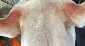
Fig. 1: Erythema in axillae in atopic dermatitis
Source: Dr Carmen Lorente
-
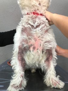
Fig. 2: Chronic atopic dermatitis with active inflammation. Reactive therapy is required.
Source: Dr Carmen Lorente
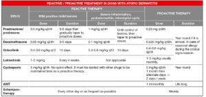
- Tab. 1: Reactive / proactive treatment in dogs with atopic Dermatitis
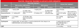
- Tab. 2: Reactive / proactive treatment in cats with atopic Dermatitis
-
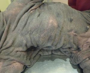
Fig. 3: Chronic atopic dermatitis with severe acanthosis. Topical therapy is needed to restructure the skin
Source: Dr Carmen Lorente
Glucocorticoids are anti-inflammatory and immunosuppressive drugs (according to the dose) that have high efficacy and rapid effect in controlling skin inflammation, pruritus and associated lesions. They can be used in dogs and cats.
For atopic dermatitis treatment, low ant-iinflammatory doses of oral corticosteroids are enough. Do not use high doses or delayed effect glucocorticoids as they are not more effective but increase the risk of side effects.
The dosage of glucocorticoids depends on their potency. Commonly used are prednisolone and prednisone. Rarely, oral triamcinolone or oral dexamethasone are used. Dexamethasone is ten times more potent than prednisolone/prednisone, so its dose is ten times lower. Side effects of glucocorticoids are proportional to drug potency, dosage and duration of administration.
Oclacitinib (Apoquel®) is an inhibitor of IL-31, the main cytokine involved in the mechanism of pruritus, by binding and blocking its receptor Janus kinase. Oclacitinib reduces inflammation and pruritus. Its effect is fast and last a maximum of 24 hours.
It is not licensed for use in cats, but there is good scientific evidence of its efficacy at a dose of 1 mg/kg every 12 or 24 hours. Its off-label use needs close monitoring of these animals.
Lokivetmab (Cytopoint®) is a caninized anti-L31 monoclonal antibody (MAb) that specifically binds to and neutralizes IL31. It is administered subcutaneously at a dose of 1-2 mg/kg and lasts for an average of 4 weeks. It has a rapid anti-pruritic effect, generally within 8 hours of its administration. As it is a species-specific MAb, it can only be used in dogs.
Cyclosporin (CsA) is an immunomodulatory, anti-pruritic, and anti-inflammatory drug. It has no immediate effect and may take up to a month to effectively control atopic dermatitis. Its most frequent side effects are gastrointestinal disorders that are normally temporary. The administration of the capsules frozen usually avoid these effects.
It is registered for cats and dogs. It is administered daily until effect, usually one month, and subsequently, the administration schedule is prolonged (alternate days -> 3-2 days per week).
Others
Today, it is known that histamine has no principal role as a mediator of pruritus in animals with atopic dermatitis. Scientific evidence shows that the efficacy of antihistamines for the treatment of AD is nil to moderate, being even less as monotherapy.
Topical treatment
Atopic dermatitis clinical picture appears differently in each animal. Individuality makes each individual have areas (ears, paws, axilla, abdomen, neck, inguinal region…) where inflammation and pruritus are more intense and flare easier. Topical treatment complements systemic treatment to control the most sensitive areas and is very useful as reactive and proactive therapy. Glucocorticoids and topical tacrolimus are effective in controlling localised puritus. The recommended glucocorticoid is hydrocortisone aceponate, as it is metabolised in the skin and has no systemic effects. Other glucocorticoids can contribute to the development of iatrogenic Cushing and skin atrophy.
3. Restructuring and hydration of the skin
Changes in the skin and the cutaneous barrier have been demonstrated in atopic animals. They are due to genetic factors associated with disease and to the inflammation. Skin changes favour the multiplication of infectious agents (bacteria and yeasts), the penetration of allergens, pruritus and predispose to the development of more skin lesions.
Topical treatment focused on restoring the skin physiology and function is crucial in atopic animals.
Shampoo therapy removes allergens deposited on the skin or hair, as well as scabs, cellular debris, secretions and bacterial agents; it helps skin restructuring and hydration and has a calming, anti-inflammatory and antipruritic effect.
In addition to baths, there are products in the form of foams, creams or spot-ons with restructuring properties.
A complete and balanced diet with nutritional complexes adapted to atopic disease is also necessary.
4. Secondary infections control
Secondary bacterial (pyoderma) or Malassezia infections are very common in atopic animals. Skin infection causes puritus and increases inflammation. Puritus due to infection is not controlled with antipruritic drugs.
In any atopic animal with active lesions, the possible existence of an infectious component must be evaluated and, if necessary, treated.
5. Allergen-specific immunotherapy (ASIT)
It is the only treatment that can reverse this incurable disease. It is a long-term treatment whose objective is to ”educate” the immune system not to overreact to environmental allergens. Its use is recommended in all animals diagnosed with atopic dermatitis.
ASIT does not have an immediate effect, as ”education” of the immune system takes time, and should be joined to the antipruritic treatment until maximal effect. Some animals can have an excellent response in 4-6 months, but the maximum response to ASIT can take 1-2 years in other cases. A recent study describes an improvement in the clinical picture in a mean time of 4.7 months. 58% of dogs were maintained exclusively with ASIT, without the need for additional medication, in less than ten months of the start of treatment.
It should be used together with antipruritic treatment until possible to reduce or suspend antipruritic treatment (better efficacy after one year of treatment).
Dr Carmen Lorente, DVM, PhD, DipECVD EBVS® European Specialist in Veterinary Dermatology
Recommended lectures:
-
Fischer NM and Müller Allergen Specific Immunotherapy in Canine Atopic Dermatitis: an Update. 2019. Current Dermatology Reports. 8: 297–302
-
Mueller RS et Treatment of the feline atopic syndrome – a systematic review. 2021. Veterinary Dermatology. 32 (1) 43-48.
-
Olivry T et Treatment of canine atopic dermatitis: 2010 clinical practice guidelines from the International Task Force on Canine Atopic Dermatitis. 2010. Veterinary Dermatology 21 (3) 233-48
-
Olivry T et Treatment of canine atopic dermatitis: 2015 updated guidelines from the International Committee on Allergic Diseases of Animals (ICADA). 2015. BMC Veterinary Research 11(1):210.
-
Olivry T, Banovic Treatment of canine atopic dermatitis: time to revise our strategy? 2019,. Vet Derm Volume30 (2) 87-90.
-
Ramió‐Lluch L et al. Efficacy of allergen‐specific immunotherapy in dogs with atopic dermatitis: a retrospective study of 145 Vet Rec.2020 Dec 19;187(12):493.
-
Tamamoto-Mochizuki C et al. Proactive maintenance therapy of canine atopic dermatitis with the anti-IL-31 lokivetmab. Can a monoclonal antibody blocking a single cytokine prevent allergy flares? Vet 2019 30. 98-e26
